Why Choosing the Best Neurosurgeon in Bangalore is Critical for Brain and Spine Health
Finding the best neurosurgeon in Bangalore is essential for anyone facing neurological conditions affecting the brain or spine. The complexity of neurosurgical procedures requires precision, expertise, and access to advanced medical technology to ensure the best possible outcomes. Without proper treatment, conditions such as brain tumors, spinal disorders, and nerve damage can significantly impact a patient’s quality of life.
A highly skilled neurosurgeon can accurately diagnose neurological disorders and recommend the most effective treatment plan. Their expertise reduces surgical risks, improves recovery rates, and enhances long-term health benefits. Choosing a qualified professional is crucial to prevent complications and ensure a smooth recovery.
Hospitals with dedicated neurosurgical teams provide comprehensive care, from diagnosis to post-surgical rehabilitation. When selecting a neurosurgeon, factors such as experience, specialization, patient reviews, and hospital facilities should be carefully evaluated. The right choice can lead to a successful treatment journey with minimal complications.
How Neurosurgery Improves Quality of Life: What You Need to Know
A neurosurgeon is a highly specialized doctor trained to diagnose and treat disorders affecting the brain, spine, and nervous system. They perform complex procedures such as brain tumor removal, spinal surgeries, and nerve decompression to improve patient outcomes. Their expertise is essential in managing conditions that impact mobility, cognition, and overall neurological function.
Neurosurgeons undergo extensive training, including medical school, residency, and specialized fellowships, to master intricate surgical techniques. Their work involves not only performing surgeries but also providing non-surgical treatments such as pain management and rehabilitation. Patients often consult them for chronic conditions like herniated discs, strokes, and epilepsy.
With advancements in minimally invasive neurosurgery, procedures have become safer, leading to faster recovery times and reduced complications. Choosing the best neurosurgeon in Bangalore ensures access to cutting-edge techniques that enhance precision and patient safety.
Neurosurgeon vs. Neurologist: Who Should You Consult?
Many patients are unsure whether they need a neurosurgeon or a neurologist for their condition. While both specialize in treating nervous system disorders, their roles are distinct. A neurologist focuses on diagnosing and managing neurological diseases without surgery, using medications, lifestyle changes, and therapies.
On the other hand, a neurosurgeon is trained to perform surgical interventions for conditions that cannot be managed with medication alone. For example, a neurologist may treat migraines, Parkinson’s disease, or multiple sclerosis, whereas a neurosurgeon handles brain tumors, spine deformities, and nerve compression issues.
If a patient is experiencing chronic pain, mobility issues, or worsening neurological symptoms, consulting a neurosurgeon may be the best option. For diagnosis and initial treatment, neurologists and neurosurgeons often work together to provide comprehensive care.
Signs You Need a Neurosurgery Consultation in Bangalore
- Chronic Headaches & Dizziness – Persistent headaches that don’t respond to medication may indicate a neurological issue requiring further evaluation.
- Severe Back & Neck Pain – Spine-related problems like herniated discs, sciatica, or spinal stenosis often need surgical intervention.
- Numbness, Weakness, or Loss of Coordination – Symptoms like difficulty walking, numbness in limbs, or loss of motor control could be signs of nerve compression or brain disorders.
- Seizures & Unexplained Blackouts – If medication does not control epilepsy or seizures, neurosurgery may be necessary for long-term relief.
- Brain Tumor Symptoms – Blurred vision, memory loss, frequent nausea, or personality changes might indicate the need for a neurosurgical evaluation.
- Spinal Cord Injuries or Trauma – Emergency neurosurgical care is required for fractures, dislocations, or nerve damage due to accidents or injuries.
- Unsuccessful Non-Surgical Treatments – If physiotherapy, medication, or lifestyle modifications have not provided relief, a neurosurgeon can explore advanced surgical solutions.
Common Brain and Spine Conditions Treated by Neurosurgeons
Neurosurgeons specialize in diagnosing and treating a wide range of brain and spine disorders that affect neurological function, mobility, and overall health. Below are the key conditions that require expert neurosurgical care.
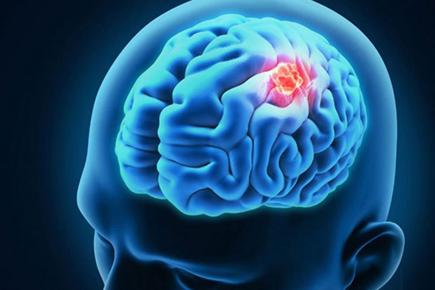
Brain Surgery: Critical Conditions That Require Neurosurgical Treatment
-
Brain Tumors
-
Abnormal cell growth in the brain, which may be benign or malignant.
-
Symptoms: Headaches, vision issues, memory loss, seizures.
-
Treatments: Surgery, radiation therapy, chemotherapy.
-
-
Stroke Management
-
Occurs due to blocked blood flow (ischemic) or a burst vessel (hemorrhagic).
-
Symptoms: Numbness, slurred speech, confusion, loss of coordination.
-
Treatments: Clot removal, aneurysm clipping, carotid artery surgery.
-
-
Epilepsy Surgery
-
For drug-resistant epilepsy with frequent seizures.
-
Surgical options: Lobectomy, vagus nerve stimulation, deep brain stimulation.
-
-
Brain Aneurysm Treatment
-
Weak blood vessels in the brain that may rupture, causing severe bleeding.
-
Symptoms: Severe headache, nausea, vision loss, unconsciousness.
-
Treatments: Aneurysm clipping or coiling.
-
-
Traumatic Brain Injury (TBI) & Skull Fractures
-
Caused by accidents, falls, or sports injuries.
-
Symptoms: Memory loss, dizziness, difficulty speaking.
-
Treatments: Craniotomy, decompressive craniectomy, brain reconstruction.
-
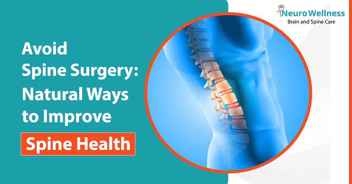
Spine Surgery: Advanced Treatments for a Pain-Free Life
-
Herniated Discs & Sciatica
-
Spinal disc bulging or rupture pressing on nerves.
-
Symptoms: Back pain, leg numbness, walking difficulty.
-
Treatment: Discectomy, spinal decompression.
-
-
Spinal Cord Injuries & Nerve Compression
-
Damage due to trauma, tumors, infections.
-
Symptoms: Paralysis, severe pain, bladder/bowel control loss.
-
Treatment: Spinal fusion, laminectomy, nerve decompression.
-
-
Sciatica & Chronic Lower Back Pain
-
Pressure on the sciatic nerve causing radiating leg pain.
-
Symptoms: Shooting pain, weakness, tingling in legs.
-
Treatment: Microdiscectomy, nerve root decompression.
-
-
Spinal Deformities: Scoliosis & Kyphosis
-
Abnormal spinal curvature affecting posture and movement.
-
Treatment: Spinal fusion, rod implantation, vertebral osteotomy.
-
-
Minimally Invasive Spine Surgery (MISS)
-
Small incisions for quicker recovery and less pain.
-
Common procedures: Endoscopic discectomy, laser spine surgery, robotic-assisted spinal fusion.
Why Neurosurgical Treatment is Essential for These Conditions
- Early intervention can prevent complications like permanent nerve damage, paralysis, or cognitive decline.
- Minimally invasive techniques reduce recovery time and post-surgical discomfort.
- Choosing the best neurosurgeon in Bangalore ensures access to cutting-edge treatments and expert care.
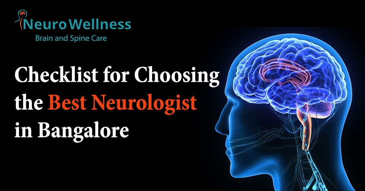
How to Choose the Best Neurosurgeon in Bangalore
Selecting the best neurosurgeon in Bangalore is crucial for effective treatment and long-term recovery. The following key factors will help you make an informed decision when choosing a neurosurgical specialist.
1. Experience & Qualifications: What to Look for in a Neurosurgeon
- Board Certification & Medical Training – Ensure the neurosurgeon is certified and trained in recognized medical institutions.
- Years of Experience – Look for specialists with extensive experience in brain and spine surgeries.
- Fellowships & Specializations – Neurosurgeons with specialized training in areas like brain tumors, spine disorders, or minimally invasive techniques often provide better outcomes.
2. Specializations: Understanding Expertise in Brain vs. Spine Surgery
- Brain Neurosurgeons – Focus on procedures like brain tumor removal, stroke intervention, and epilepsy surgery.
- Spine Neurosurgeons – Specialize in conditions like herniated discs, spinal cord injuries, and deformities.
- Minimally Invasive Specialists – Use advanced techniques such as robotic-assisted surgery, laser surgery, and endoscopic spine procedures.
3. Success Rate & Reviews: How to Check Patient Testimonials and Hospital Ratings
- Patient Testimonials & Case Studies – Look for real-life success stories of patients treated by the neurosurgeon.
- Google & Hospital Reviews – Check ratings on platforms like Google, Practo, and hospital websites.
- Word-of-Mouth & Referrals – Recommendations from past patients, family, or primary doctors can be helpful.
4. Technology & Facilities: Importance of Advanced Neurosurgical Tools
- State-of-the-Art Equipment – Ensure the hospital has modern tools like MRI-guided surgery, neuronavigation, and robotic surgery.
- Availability of ICU & Emergency Care – Advanced post-operative care is essential for neurosurgery patients.
- Access to Multidisciplinary Teams – A team approach involving neurologists, physiotherapists, and rehabilitation specialists enhances recovery.
5. Hospital Accreditation: Why Choosing the Right Hospital Matters
- JCI & NABH Accreditation – Certified hospitals maintain international healthcare standards.
- Infrastructure & Patient Care – A well-equipped hospital improves the success rate of neurosurgical procedures.
- Post-Surgical Support – A hospital that provides rehabilitation, physiotherapy, and follow-up care ensures better long-term results.
Checklist for Choosing the Best Neurosurgeon in Bangalore
✔ Does the neurosurgeon have board certification and advanced training?
✔ Does the specialist have a high success rate for complex neurosurgical procedures?
✔ Are there positive patient reviews and testimonials?
✔ Is the hospital equipped with modern neurosurgical technology?
✔ Does the hospital provide comprehensive post-surgical care and rehabilitation?
Choosing the right neurosurgeon can make a significant difference in your treatment and recovery journey. Take time to evaluate your options and select a specialist who aligns with your healthcare needs.
Best Neurosurgery Hospitals in Bangalore
Choosing the right hospital is just as important as selecting the best neurosurgeon in Bangalore. The hospital’s infrastructure, technology, and multidisciplinary team play a crucial role in ensuring successful neurosurgical outcomes.
Why Choose NeuroWellness Brain and Spine Hospital?
✔ Top-rated neurosurgeons in Bangalore with years of expertise.
✔ Cutting-edge neurosurgical technology for safe and effective treatment.
✔ Personalized treatment plans for brain and spine conditions.
✔ Post-surgical care and physiotherapy to ensure complete recovery.
✔ High success rates in complex neurosurgical procedures.
Conclusion
Choosing the best neurosurgeon in Bangalore is crucial for receiving high-quality care for brain and spine conditions. The right specialist, combined with an advanced medical facility, can significantly improve patient outcomes and recovery rates. Whether it’s a complex brain tumor surgery, spine treatment, or minimally invasive neurosurgical procedure, selecting an experienced neurosurgeon ensures precision, safety, and optimal results.
Frequently Asked Questions (FAQs)
1. How do I choose a neurosurgeon?
Choosing the best neurosurgeon requires careful evaluation of the following factors:
✔ Board Certification & Experience – Ensure they are NABH-certified and have performed similar surgeries.
✔ Patient Reviews & Success Rates – Look for Google reviews and hospital ratings.
✔ Hospital Facilities – Choose hospitals with MRI-guided surgery and neuronavigation.
✔ Consult Multiple Experts – Always get a second opinion before major surgeries.
2. What is the best hospital for neurosurgeons?
The best hospitals for neurosurgery in Bangalore include:
✔ NeuroWellness Brain & Spine Hospital@Kauvery hospital – Specialized in brain tumor surgery & minimally invasive spine surgery.
✔ Manipal Hospitals – Known for neuro-oncology and stroke management.
✔ Aster CMI Hospital – Offers advanced robotic-assisted neurosurgery.
✔ Apollo Hospitals – Best for deep brain stimulation & skull base surgery.
3. What are the most common brain and spine surgeries performed by neurosurgeons?
Neurosurgeons perform various brain and spine surgeries to treat neurological disorders, including:
✔ Brain Tumor Surgery – Removal of malignant or benign tumors affecting brain function.
✔ Spinal Decompression Surgery – Relieves pressure on spinal nerves caused by herniated discs.
✔ Stroke Surgery – Procedures such as clot removal and aneurysm repair to prevent brain damage.
✔ Epilepsy Surgery – Reduces seizure frequency in patients with drug-resistant epilepsy.
✔ Minimally Invasive Spine Surgery (MISS) – Advanced procedures for quicker recovery with less scarring.
4. When should I see a neurosurgeon instead of a neurologist?
While neurologists treat conditions with medications and therapy, neurosurgeons handle cases requiring surgical intervention. You should consult a neurosurgeon if you experience:
✔ Chronic back or neck pain unresponsive to physical therapy.
✔ Severe or frequent headaches that indicate brain abnormalities.
✔ Weakness, numbness, or loss of movement due to spinal cord compression.
✔ Brain tumors, aneurysms, or severe head injuries requiring immediate surgery.
✔ Failed non-surgical treatments for epilepsy, spine disorders, or nerve compression.
5. What should I expect during my first consultation with a neurosurgeon?
Your first consultation with a neurosurgeon will include:
✔ Medical history review – Discussion of symptoms, past treatments, and family history of neurological disorders.
✔ Diagnostic tests – MRI, CT scans, or nerve conduction studies may be recommended.
✔ Treatment options – The neurosurgeon will explain whether surgery is required or if alternative treatments are available.
✔ Surgery discussion (if needed) – Risks, recovery process, and expected outcomes of surgical procedures.
✔ Post-consultation plan – Further tests, second opinions, or scheduling surgery if necessary.