Brain aneurysm clipping is a surgical procedure to prevent rupture by sealing off a weakened blood vessel in the brain. Recovery includes rest, managing fatigue, and follow-up care.
Consult Neurowellness if you’ve had headaches, visual changes, or were diagnosed with an unruptured aneurysm.
Brain Aneurysm Clipping: A Comprehensive Guide
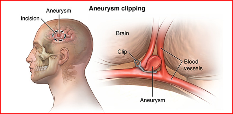
Brain Aneurysm Clipping is a procedure for treating an aneurysm. It is a balloon-like bulging in the artery wall. Aneurysms wall will be very thin that they can leak or rupture, discharging blood into the brain’s surrounding areas. A neurosurgeon performs a craniotomy, which involves opening the skull and placing a tiny clip across the aneurysm’s neck to stop or prevent bleeding.
What is aneurysm clipping?
Surgical clipping is used to isolate an aneurysm from the rest of the body’s circulation without obstructing any neighbouring very important small vessels called perforating arteries. Perforating arteries give blood supply to very important brain centres. A craniotomy is a procedure that involves making a hole in the skull and remove the part of the bone under general anaesthesia. To find the aneurysm, the brain is softly pulled. To prevent normal blood flow from entering the aneurysm, a tiny clip is put across the base of the aneurysm neck.
The clip functions similarly to a miniature coil spring clothespin, with the blades remaining tightly closed until pressure is applied to open them. The titanium clips are permanently attached to the artery.
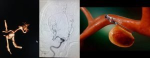
Aneurysms will appear on angiogram in different of sizes and shapes- blister, small, large and giant. At their origin on the main artery, saccular aneurysms have a neck and a dome that can expand and grow like a balloon. Clips are easier to lay across these. Some aneurysms have a broad neck, while others are fusiform in appearance with no discernible neck. It’s harder to clip across these.
The following conditions may benefit from clipping:
・ Ruptured aneurysms
A subarachnoid haemorrhage occurs when a aneurysm is located in the blood vessel between the brain and the skull bursts open and releases blood into the space between the brain and the skull (SAH). Within the first 14 days after the initial bleed, there is a 22% chance of bleeding again. As a result, surgery must be scheduled within 72 hours following the initial bleed or as early as possible.
・Unruptured aneurysms
They are usually discovered during normal testing for another ailment and do not produce symptoms. Aneurysm rupture occurs roughly once every year, however the risk varies based on the size and location of the aneurysm. When a rupture occurs however the risk of death is 40% and the chance of disability is 80%.
After addressing the risks and benefits of aneurysm clipping, a brain aneurysm is detected using neuro imaging DSA or CT angiography.
If aneurysm is giant, brain bypass surgery may be required to establish the blood flow into the artery after clipping. Bypass surgery is similar to a heart bypass surgery. One of the blood vessel is taken or harvested from above the scalp or retrieved from a limb and linked to the brain vessel with very thin suture material in a brain bypass technique.
Procedure :
・ Who performs the procedure?
A skilled Neurosurgeon in Bangalore, who is trained in cerebro vascular neurosurgery performs the surgical clipping. Cerebrovascular surgery is a sub speciality of neurosurgery. Inquire about your surgeon’s training, especially if your situation is complicated.
・What happens before surgery?
If you have been brought to the emergency room with a ruptured aneurysm, you will need to prepare differently than if you are having an unruptured aneurysm clipped.
A ruptured aneurysm is life-threatening; you may be rushed to the operating room right away after the doctors have discovered it and stabilised your blood pressure and based on your clinical status.
・Top neurologists in Bangalore utilize medicine, hyperventilation using a breathing machine or sedative to stabilise blood pressure. To monitor blood pressure, an arterial pressure line may be put into your arm. Visits with your loved one may be limited in order to maintain a peaceful and unstimulating environment.
・You will fill out paperwork and sign consent forms if you have an unruptured aneurysm so that your surgeon is aware of your medical history. Preoperative tests, e.g, electrocardiograms and chest X rays may be required several days prior to surgery.
・ Seven days before surgery stop taking all non steroidal anti inflammatory drugs and blood thinners. To avoid bleeding and healing issues, stop using nicotine and drinking alcohol one week before and two weeks after the surgery.
What happens during surgery?
This procedure follows six steps which are as follows:
Step 1: Prepare the patient
・You will be sedated and given general anaesthesia while lying on the operating table.
・Your head is placed in a three-pin skull fixation device after you have fallen unconcious, which attaches to the table and keeps your head in place during surgery. The scalp’s incision region is next prepared.
・ Only 1/4th inch broad area along the anticipated incision is shaved using a hair sparing procedure. The entire incision region may be shaved in some cases.
・Mannitol, a brain relaxant may be prescribed.
Step 2: Perform a craniotomy
・A bone flap or craniotomy will be formed in your skull depending on the location of the aneurysm. Inquire with your surgeon about the specific location of the skin incision and the bone removal .
・The surgeon will make a skin incision to expose the skull after your scalp has been prepped. A drill is then used to make burr holes in the skull.
・The dura mater or protective covering of the brain, is exposed by lifting and removing the sliced bone flap. The bone flap is placed in a safe area and will be restored at the conclusion of the treatment.
Step 3: expose the aneurysm
・The brain is exposed after the dura is opened. To gently open a corridor between the brain and the skull, retractors can be used.
・The surgeons carefully locates the artery and traces it to the aneurysm using an operating microscope.
・Before putting the clip, the surgeon establishes control of the blood flow in and out of the aneurysm. Handling of the aneurysm, especially the dome, can trigger rupture.
Step 4: insert the clip
・The neck of the aneurysm is prepared for clipping. Connective tissue often holds it in place, so it must be freed and isolated from other structures.
・Small arteries called perforators must be noticed so they are not included in the clip.
・When the clip is released, the jaws close, squeezing the aneurysm from the parent artery. It is possible to use multiple clips.
Step 5: check the clip
・The surgeon examines the clip to ensure that it is not limiting the parent artery or that it has other arteries in its jaws.
・To confirm blood flow through the parent artery, intraoperative angiography(ICG) may be used
Step 6: close the craniotomy
・Sutures are used to close the dura. The bone flap is replaced and titanium plates and screws are used to fix it to the skull
・Sutures are used to reattach the muscles and skin.
・Over the incision, a soft adhesive dressing will be applied
What happens after the surgery?
・You will be brought to the recovery room after surgery, where your vital signs will be monitored as you wake up from anaesthesia.
・ After that, you will be moved to an intensive care unit (ICU) for observation and monitoring.
・As needed, pain medication will be administered.
・Following surgery you may have nausea and headaches. These symptoms can be managed with medication.
Summary
Patients with aneurysms may experience short and long term problems as a result of a rupture or treatment. With healing and counselling some of these deficiencies may fade away. The rehabilitation procedure for burst aneurysms can take months or years to appreciate the impairments you have suffered and regain function.
“Clipping offers long-term protection against aneurysm rupture. While it’s a complex surgery, in the right hands, it can be life-saving.”
— Dr. Ganesh Veerabhadraiah
Learn More:
endovascular coiling of superior cerebellar aneurysm
top 7 things you need to know about transient ischemic attack tia stroke treatment inbangalore