Neurosurgical emergencies are very important. Any delay in treating may influence patients outcome. Early diagnosis and treatment is very important, which can save life and prevent brain and spinal cord damage further.

Neurosurgical emergencies are very important. Any delay in treating may influence patients outcome. Early diagnosis and treatment is very important, which can save life and prevent brain and spinal cord damage further.
Spine surgery is done to repair the disc! – No, Spine Surgery is not done to repair the damaged disc, but to relieve the pain and prevent neurological deficits.

6 out of 7 billion people own a cell phone which is pretty shocking as to the fact that only 4.5 billion people have access to a working toilet.!!!

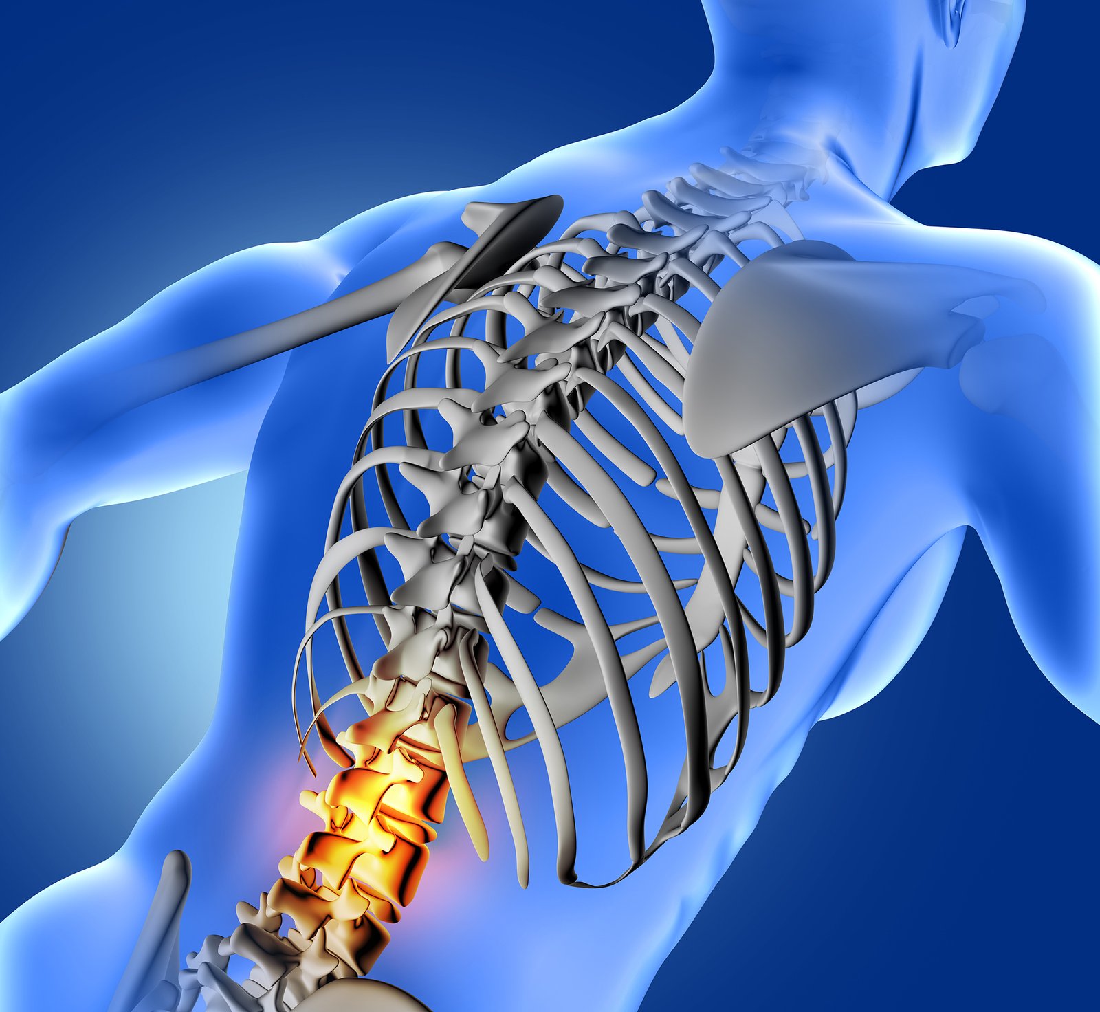
Back pain is a common neurological complaint noted in our out patient department. It could be acute and chronic.

When mild and moderate pain has become very severe, It is radiating to one of the leg, Developing weakness in the feet or toes and unable to wear slipper,
On coughing and sneezing or yawning…

Muscle strain or ligament strain occurs due to twisting of back, lifting heavy weights, sudden jerky movements, abnormal posture for long time. Radicular pain due to lumbar (PIVD) disc prolapse or disc bulge.

Find out the best comfortable position for you to sleep. Moderate size pillow for neck (very thin or very thick pillow would cause strain on neck). Get a good mattress – soft preferably, there is no need to go for very expensive mattress.