What are the risk factors of stroke we should know?

A stroke can However, if you have specific risk factors, your chances of having a stroke increase. Some risk factors of stroke can be altered or managed, while others cannot. Today, an increasing number of people are putting their most valuable asset their brains under protection. Are you one of them?
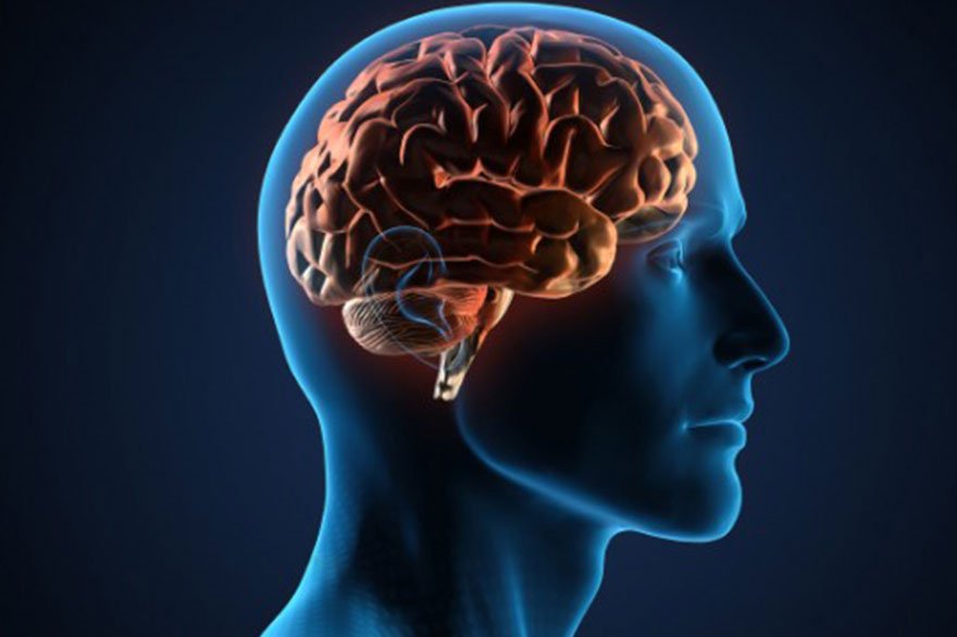
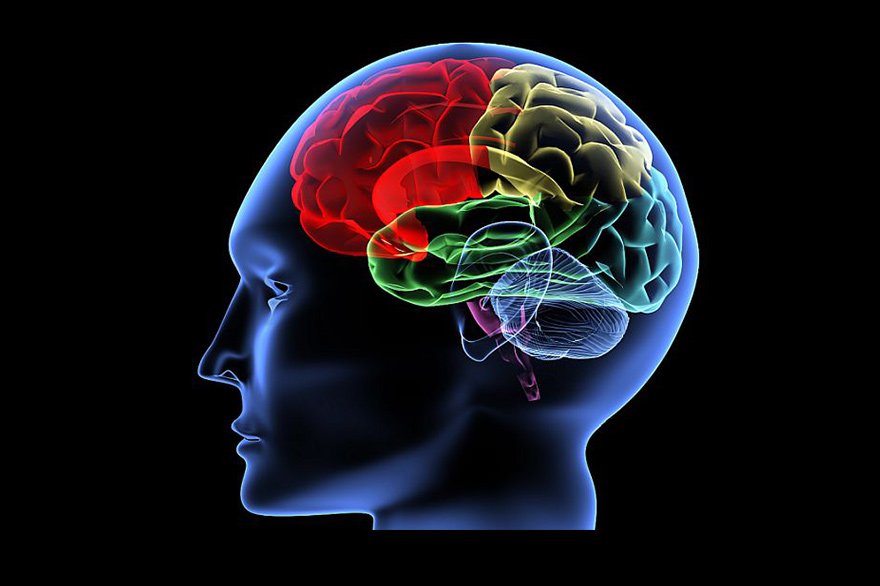
What are the risk factors of stroke that cannot be modified:

1. a previous stroke or pre-existing cardiovascular disease such as a heart attack.

2. Age: 60 years old or older.

3. Family history: Members in the family that have suffered a stroke

4. Gender: Males are at a higher risk than females.

5. Race: Black, Asian, and Hispanic

6. Sickle cell disease, polycythemia, protein C/S deficiency, hyperhomocysteinemia, etc., are blood diseases that cause excessive clotting.

7. Mitral stenosis (a type of valvular disease)

8. Genetics or heredity: People with a family history of stroke have a higher risk of having a stroke.

What are the risk factors of stroke that can be modified: 

Risk factors of stroke that can be altered, treated, or controlled medically include

1. High blood pressure: persistent Blood pressure of 140/90 or above can cause damage to the brain’s blood vessels (arteries).

2. Heart disease: There is a strong association between heart disease and stroke. Several types of cardiac disease are known to increase the risk of stroke. Stroke, like coronary heart disease, is a risk factor. Atherosclerosis (hardening of the arteries) increases the risk of stroke in people with coronary heart disease, angina, or who have had a heart attack.

3. Diabetes: Control your blood sugar if you have Type 1 or Type 2 diabetes. Diabetes mellitus is a risk factor for stroke on its own. Many diabetics also have high blood pressure, high cholesterol, and are overweight, all of which increase their risk. Even though diabetes is curable, it still raises your risk of stroke.

4. Smoking: Cigarette smoke contains nicotine and carbon monoxide, which harms the cardiovascular system and increases the risk of stroke. When birth control tablets are taken with cigarette smoking, the risk of stroke is considerably increased.

5. History of TIAs:  Mini-strokes is a term used to describe TIAs. The symptoms are similar to those of a stroke, although they don’t stay as long. You’re almost ten times more likely to suffer a stroke if you’ve had one or more TIAs than someone your age and sex who hasn’t.

6. High red blood cell count: The blood thickens and clots are more likely when the quantity of red blood cells increases significantly. This increases the chances of having a stroke.

7. High blood cholesterol and lipids: High cholesterol levels can contribute to artery thickening or hardening (atherosclerosis), which is caused by plaque buildup. Plaque is a buildup of fatty substances, cholesterol, and calcium in the arteries. The amount of blood flow to the brain can be reduced by plaque accumulation on the inside of the arterial walls. When the brain’s blood supply is cut off, a stroke develops.

8. Lack of physical activity

9. Obesity

10. Excessive alcohol consumption: Blood pressure rises if you drink more than two drinks every day. Stroke can occur as a result of binge drinking.

11. Illegal drugs: Abuse of intravenous (IV) drugs increases the risk of a stroke due to blood clots (cerebral embolisms). Cocaine and other narcotics have been linked to heart attacks, strokes, and a variety of other cardiovascular issues.

12. Abnormal heart rhythm: Some types of heart disease can increase your chances of having a stroke. The most potent and modifiable heart risk factor for stroke is having an abnormal heartbeat (atrial fibrillation).

13. Cardiac structural abnormalities: Long-term (chronic) heart damage can be caused by damaged heart valves (valvular heart disease). This can increase your risk of stroke over time.

Other risk factors of the stroke to consider are:

Where you reside: Strokes are more common in the southeast than in other parts of the country. This could be due to variances in lifestyle, race, smoking habits, and diet between regions.

Temperature, season, and climate:  Stroke deaths are more common during periods of excessive heat.

Social and economic factors: Strokes are more likely in low-income people, according to some studies.

Neurowellness provides high-quality Advanced Stroke treatment in Bangalore at an affordable cost in comparison with the other medical treatment options worldwide.

What is spondylolisthesis?

Lower back pain may be caused by spondylolisthesis, a spinal disorder. It happens when one of your vertebrae or spine bones slips out of place and lands on the vertebra below it. Nonsurgical treatment can usually alleviate your problems. In most cases, surgery for severe grade spondylolisthesis is effective.

What is spondylolisthesis?

Spondylolsthesis is a condition in which the vertebrae move more than they should, resulting in spine instability. A vertebra slips out of place and lands on the one below it. There will be pars interarticularis defect a bone connecting upper and lower facet. It could exert pressure on a nerve, resulting in lower back or leg pain.

The Greek words spondylos, which means “spine” or “vertebra” and listhesis, which means “slipping, sliding or moving” are combined to form spondylolisthesis (pronounced sphon-di-low-less-THEE-sis).

Types of spondylolisthesis:

  •  Degenerative spondylolisthesis: the most prevalent kind is degenerative spondylolisthesis, which occurs as a result of ageing.
  • Isthmic spondylolisthesis: as a result of spondylolisthesis, isthmic spondylolistheis develops. The bone is weakened by the crack or fracture.
  • Congenital spondylolisthesis: when a baby’s spine does not form properly before birth, this is known as congenital spondylolisthesis. The person’s vertebrae are misaligned, putting them at risk for slippage later in life.
  • Less common types of spondylolisthesis are:
  1. Traumatic spondylolisthesis: it occurs when the vertebrae slip as a result of an injury.
  2. Pathological spondylolisthesis: when a disease, such as osteoporosis, or a tumour, develops pathological spondylolisthesis.
  3. Post surgical spondylolisthesis: slippage as a result of spinal surgery is known as post-surgical spondylolisthesis.

How common is spondylolisthesis?

Spondylolisthesis and spondylolysis affect approximately 4% to 6% of the adult population. It is easy to go years without realising you have spondylolisthesis because you do not have any symptoms.

Degenerative spondylolisthesis (which happens as a result of ageing and wear and tear of the spine) is more common in women than in men after the age of 50.

Isthmic spondylolisthesis (typically induced by spondylolysis) is one of the most common reasons for back pain in teenagers.

Causes of spondylolisthesis:

One of the most common causes of spondylolisthesis in young athletes is overextending the spine. Genetics could also have a role. Some people are born with spinal bones that are thinner than others. Wear and tear on the spine and discs (the cushions between the vertebrae) can develop this problem in older persons.

What are the symptoms of spondylolisthesis?

You might not notice any signs or symptoms of spondylolisthesis. Some people are unaware that they have the disease. If you do experience symptoms, the most common one is lower back ache. The discomfort may spread to the buttocks and thighs. You may also experience:

  • Back pain/leg pain aggravated by activity- working at home or bending and picking up object.
  • Stiffness in the back
  • Spasms in the hamstring muscle (muscles in the back of the thighs)
  • Walking or standing for long periods of time is difficult
  • When leaning over, there is pain
  • Numbness, tingling or weakness in the foot.

How is spondylolisthesis diagnosed?

Your doctor will conduct a physical examination and inquire about your symptoms. An imaging/scan will very certainly be required to confirm the diagnosis.

What imaging tests will be needed?

  • A spinal X ray allows doctors to see if a vertebrae is misaligned.
  • To see the spine in greater detail or to see soft tissue like discs and nerves, a CT scan or an MRI scan may be required.

How to reduce the risk of spondylolisthesis?

You can lower your chance of spondylolisthesis by doing the following:

  • Regularly exercise your back and abdominal muscles to keep them strong
  • Maintain a healthy body mass index (BMI). Weight gain puts additional strain on your lower back
  • To keep your bones well nourished and robust, eat a well balanced diet.

What is the outlook for people with spondylolisthesis?

 The success rate of surgery is very high. Spondylolisthesis surgery patients typically return to an active lifestyle after a few months following surgery. Following surgery, you will almost certainly require therapy to help you regain full function.

Summary:

Spondylolisthesis is one of the  common cause of back pain /discomfort, however it is not life threatening. There are a variety of therapists available, ranging from medicine to physical therapy to spinal surgery. If you are experiencing low back discomfort or finding it difficult to walk, stand, or bend over, consult the Neuro Wellness Care Center, they will provide the best and cost friendly Back Pain Treatment in Bangalore.

Neuro Endovascular Coiling

In this blog, we are throwing some lights on “Neuro Endovascular Coiling”. You will get to know all those important details of Neuro Endovascular coiling that will help you take better decisions about discussing the risks and benefits of aneurysm coiling procedures with a Neurosurgeon in Bangalore.

What is Aneurysm?

An aneurysm in the brain is a weak area in an artery in the brain that bulges out and fills with blood. In some of the cases, it balloons out of the thinned vessel wall can lead to dangerous complications if ruptures.

Once brain aneurysm is diagnosed by neuroimaging DSA or CT angiogram, after discussing benefits and risks of coiling of aneurysm coiling procedures undertaken. One should discuss the risks and benefits with a doctor.

Usually coiling procedure is suitable for aneurysms in posterior circulation that is vessels supplying the brain stem and cerebellum, narrow neck of aneurysm. You should always discuss with neurosurgeons about the coiling and other approaches, benefits and risks.

Endovascular coiling is usually carried out under general anaesthesia in a cath lab.

It is usually done from an artery in the groin called the femoral artery. Other arteries can be radial or brachial arteries in hand. Here catheters and micro guide wires are used to reach the area of the aneurysm.

Platinum coils are used to fill the aneurysm and seal that prevent the rupture. Sometimes additionally balloons or stents are used to facilitate the filling of platinum coils.

How you should be prepared for an endovascular coiling?

  • In order to do the procedure, you will be asked to read the consent form carefully and sign it as well
  • Share all your information regarding your reactions or  allergies to alternative dyes or iodine
  • Do follow the advisories given by the doctor before surgery
  • The news regarding pregnancy shall be shared
  • Blood tests are necessary so as to see how much time does it take to clotting of blood
  • Tell the doctor about the medicines and supplementary you have been taking

Endovascular Coiling Procedure:

A coiling procedure will require you to stay in the hospital. Procedures may differ depending on your situation and the procedures of your Neurosurgeon or practitioner.

  1. Any clothing, jewellery, hairpins, dentures or anything that could interfere with the treatment will be asked to remove but generally, a hospital gown will be provided to you
  2. Before the treatment begins, you will be given time to empty your bladder
  3. On the X-ray table, you will be positioned on your back
  4. In your hand or arm, an IV (intravenous) line will be started
  5. An electrocardiogram (ECG) monitor will be attached to you, which will record your heart’s electrical activity
  6. During the process, you will have your heart rate, blood pressure, breathing rate and nervous system indication checked
  7. To drain your bladder, a catheter may be inserted
  8. Your pulses may be checked below the groyne area where the catheter will be put by the radiologist or neurosurgeon
  9.  A guide will be used to insert a catheter into the artery in your groyne
  10.  Fluoroscopy will be used to guide the catheter through the blood artery into the brain
  11.  This is a sort of X-ray that resembles an X-ray movie
  12.  Contrast dye will be given after the catheter reaches the afflicted artery in the brain, allowing the aneurysm and surrounding blood vessels to be seen on X-ray
  13.  The aneurysm will be measured and its shape and other characteristics will be recorded by the provider
  14.  The initial catheter will be replaced with a smaller catheter by the physician
  15.  The provider will insert the coil into the aneurysm once the catheter has reached it
  16.  The coil is removed from the catheter once it has been entirely inserted into the aneurysm
  17.  The doctor will insert as many coils as necessary to seal the aneurysm
  18.  Inside the aneurysm, the coils will form a mesh-like structure
  19.  The catheter will be removed once the aneurysm has sealed. In the end, the dressing will be done

There are certain risks present in this neurosurgical procedure so always consult the Best Neurosurgeon in Bangalore. There have been cases of bleeding in an aneurysm and recanalization. Endovascular coiling may not be a complete success in all the patients.

When the procedure is going on, complications such as aneurysm rupture or stroke may arise. Aneurysms can be prevented from rupturing by preventing blood flow into them.

Other risks involved are:

  • Bleeding
  • Blood clot
  • Infection
  • Rupture of aneurysm
  • Loss of consciousness
  • Paralysis on half of the body
  • Transient ischemic attack

Endovascular coiling is a minimally invasive surgery, there no craniotomy or cutting of bone, no opening of the brain, no retraction of brain or handling of blood vessels, get discharged faster and going back to normal life is fast. At NeuroWellness you will get the best and cost-effective treatment for any brain or spine related problem from the Best Neurosurgeon in Bangalore.

Neuro endovascular coiling offers a safe, less invasive solution to treat brain aneurysms. Timely diagnosis and expert care can save lives.”
Dr. Ganesh Veerabhadraiah

FAQs on Neuro Endovascular Coiling

  • Q1: What is neuro endovascular coiling?
    It is a minimally invasive procedure used to treat brain aneurysms by placing coils inside the aneurysm to block blood flow.

  • Q2: Is coiling better than open surgery for aneurysms?
    Yes, for many patients, coiling is safer with quicker recovery, but eligibility depends on aneurysm type and location.

  • Q3: How long does it take to recover from coiling?
    Most patients recover within 1–2 weeks. Some may need longer observation depending on individual health.

  • Q4: Is neuro coiling available in Bangalore?
    Yes, advanced neuro centers like NeuroWellness offer expert coiling procedures by trained neuro-interventionists.


Concussive Head Injury

  1. WE WILL KNOW ABOUT WHAT IS CONCUSSIVE HEAD INJURY?
  2. SYMPTOMS OF CONCUSSIVE HEAD INJURY?
  3. WHAT SYMPTOMS YOU SHOULD NOT NEGLECT EVEN AFTER CONCUSSION!
  4. COMPLICATIONS OF CONCUSSIVE HEAD INJURY.
  5. MANAGEMENT OF HEAD INJURY.
  6. SPORTS AND CONCUSSION.
  7. GENERAL TIPS OF CONCUSSION.

Today we will know about concussive head injury.

Concussion is a milder form of traumatic brain injury. 75% of the traumatic brain injuries include concussions. Concussions can occurs due to direct impact over the head most common reasons are – road traffic accident, fall from height, collisions or assault and sudden whiplash injury(that is sudden flexion and extension movements).

Actually concussion means SHAKE VIOLENTLY in Latin(concutere). After the impact, Brain will temporarily stop functioning.

Children also can have concussion like in adults, most common causes in them are fall from height, sports related, road traffic accidents and child abuse.

Interesting thing to note about concussive head injury is –

  • It is a low velocity trauma
  • Loss of consciousness may not be present always
  • It will not cause permanent injury
  • It is not life threatening but its symptoms need treatment with the neurosurgeon Bangalore.

Secondary Concussions are more serious and dangerous. Secondary concussion means once primary concussion already present, again one more concussion if person suffers – which may happen with boxing and wrestling where repetitive injuries are common.

1. What is concussive head Injury?

Concussive head Injury is an injury that occurs after direct trauma to head or Violent shaking of the head and upper body. This can also occur as a result of rapid acceleration-deceleration of the head as in whiplash injuries or blast injuries in war zone. There will be movement of the brain tissue within the rigid skull surrounded by the brain fluid called CSF-cerebrospinal fluid. There will be transient physiological dysfunction of the brain wherein the brain will not function for sometime, which results in symptoms.

2. What are the symptoms of concussive head injury?

  • Loss of consciousness.
  • Transient Amnesia ( memory loss ) – for a period of few minutes to hours there will be memory loss. May occur before the impact or after the impact to head. Antgade amnesia and retrograde amnesia.
  • headache, Vomiting
  • Seizures – may occur immediately or few minutes to days after the impact.
  • Giddiness – injury to balancing centre can cause balancing problems and difficulty in walking.
  • Weakness in limbs.
  • Confusion
  • Visual dysfunction
  • Dizziness
  • Irritability.
  • Loss of taste and smell
  • Hypersensitivity to light

3. Can a person have normal memory after concussive head injury?

Yes, even though few people can have some irritability, behavioural problems, most of the time,as time passes the person can have normal memory and most of the patients fully recover after the concussion. Even I had concussive head injury few years ago and I am normal now.

4. What are the complications of head injury /or post concussive syndrome?

  • Focusing problem/concentration problem
  • Sleep dysfunction-insomnia
  • Fatiguability
  • Excessive drowsiness for days to weeks
  • Personality changes
  • Memory disturbance.
  • Post traumatic stress syndrome.

5. What are the investigations advised by the Neurosurgeon in Bangalore?

CT/MRI

  • You will need Brain imaging – most commonly CT scan BRAIN and when CT scan is contraindicated like in pregnant women
  • MRI is advised since MRI is safe in pregnancy.
  • This will be normal in concussive head injury.

6. Management

Physical examination – assess level of consciousness, checking pupils, eye movements, power of limbs and also need to inspect for cut wound over the head and neck.

Most of the time – Require neuro monitoring, with medications including analgesics, antacids and antiemetics and vitamins. Antibiotics are advised if there is any open wound.

7. Head injury in Sports

Sports has a risk of concussion due to head injury. It is necessary to wear protective gear like helmet and Shield. Wearing helmet reduces 85% of head injury. It can occur with Baseball and softball

  • Cycling
  • Foot ball – CTE – Chronic traumatic encephalopathy may found in old foot ball players
  • Cricket
  • Hockey
  • Soccer
  • Boxing
  • Skating
  • Snow boarding
  • Wrestling
  • Horseback riding
  • Powered recreational driving
  • Bull riding
  • Martial arts

8. Even after mild head injury – if you are having any of these symptoms one should not neglect.

The symptoms include

  • People who are taking blood thinners
  • If person having Persistant headache
  • Person Becoming more irritable
  • Persisting vomiting after few days
  • Persisting memory disturbance
  • Persons who have consumed alcohol or on recreational drugs need to immediately seek medical help following injury

9. General tips

  • Supervision of younger children
  • Wearing Seat belt while in car
  • Follow the rules of swimming pool or water park
  • Please do not to dive in shallow water less than 12 feet
  • Some cloths can interfere with vision – Do not wear. wear clothes that interfere with vision
  • If your sleep is not good or if you have inadequate rest, don’t participate in sports
  • Obey traffic rules
  • Don’t wear compromised sport equipment.
  • Wear helmet
  • Do not drink and drive
  • Do not text while driving/walking
  • Don’t do wheeling – common in youngsters for fun.
  • Do not Involve in physical fights.

** this video is made for educational purpose, content and images are used. Any objections please mail us we will remove the contents.

Our expert Neuro team at Neurowellness in Bengaluru provides consultation and treatment to all neuro problems.

Please visit

Neurowellness
Brain and Spine Clinic
#1224, Ground-Floor, 26th Main, Jayanagar 9th Block
BANGALORE – 560069

Phone No
72596 69911
73490 17701

Website www.neurowellness.in

Facebook www.facebook.com/neurowellness.in/

Disclaimer: This information is not meant to be a substitute for professional medical advice. The reader is advised to always seek the advice of a physician prior to changing any treatment or to receive answers to questions regarding a specific medical condition.

    DOWNLOAD EBOOK

      DOWNLOAD EBOOK

        DOWNLOAD EBOOK

          DOWNLOAD EBOOK

            DOWNLOAD EBOOK

              DOWNLOAD EBOOK