Surgical Management of a Colloid Cyst of third ventricle
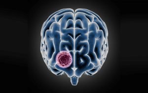
A 20-year-old girl, an art student, presented with two episodes of transient loss of consciousness lasting 20–30 minutes, accompanied by neck pain radiating to the left side. Clinical evaluation, followed by MRI, revealed a third ventricular colloid cyst, causing obstruction at the foramen of Monro and resulting in intermittent symptoms of raised intracranial pressure.

The patient underwent neuro-navigation-guided craniotomy and successful excision of the colloid cyst. Intraoperative and postoperative management were uneventful. The patient was extubated immediately following the procedure. A postoperative CT scan confirmed complete excision of the cyst, with no residual tumor visible.
She was monitored in the ICU for one day, where she demonstrated remarkable recovery. Impressively, she resumed her artistic pursuits on the first postoperative day, highlighting her swift neurological recovery. Currently, she is stable and recovering well in the ward.
We extend our gratitude to the entire neurosurgery team and the operating theater staff for their seamless coordination and dedication to achieving an excellent outcome for the patient.
This case underscores the importance of timely diagnosis, precise surgical planning, and multidisciplinary teamwork in the successful management of colloid cysts.
Histopathology- Nimhans
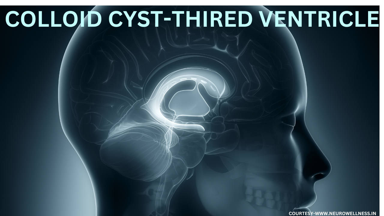
Colloid cyst

Regarding Third ventricular colloid cysts , these are rare 1 to 3 % whole intracranial tumours but potentially life-threatening intracranial lesions, often presenting a significant surgical challenge due to their deep midline location and proximity to critical neurovascular structures. These cysts can cause acute hydrocephalus and elevated intracranial pressure through obstruction of the foramen of Monro, leading to symptoms such as severe headaches, intermittent loss of consciousness, or, in rare cases, sudden death. Surgical management, either through neuro-navigation-guided craniotomy or endoscopic resection, demands meticulous planning and precise execution to minimize complications. Advances in neuro-navigation and microsurgical techniques have significantly enhanced the safety and efficacy of these procedures, making early diagnosis and prompt intervention essential in preventing catastrophic events.
Dr Ganesh Veerabhadraiah
HOD and Senior Consultant Neurosurgeon
Kauvery hospital
Electronic City
Bengaluru
Ph no 7249669911
#ColloidCyst #ThirdVentricularTumours #BrainTumours #IntraventricularTumours #EndoscopicTumourRemoval #Craniotomy