Meningitis and Amoebic Encephalitis: Symptoms, Risks & Prevention

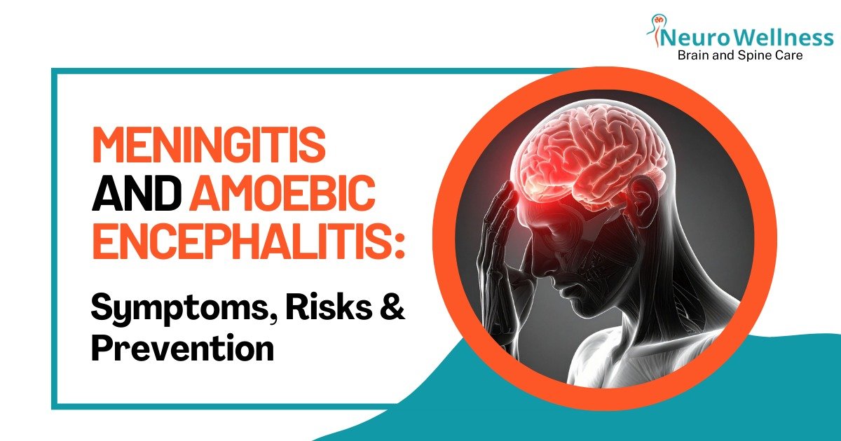
Meningitis and amoebic encephalitis are among the deadliest brain infections worldwide. While meningitis still claims lives despite treatment, amoebic encephalitis (caused by Naegleria fowleri) has a mortality rate of over 90%. Both conditions progress rapidly, making early recognition and prevention critical for survival.

In this blog, Dr. Ganesh Veerabhadraiah, senior neurosurgeon at Neurowellness, explains the symptoms, risks, and preventive steps everyone should know.

The brain is the most fragile part of the body. It works like a command center, guiding how we move, talk, breathe, and think. It is protected by a clean fluid called cerebrospinal fluid (CSF) and strong coverings called meninges.

Normally, no bacteria or parasites should ever enter this space. But if they do, the results can be deadly.

Encephalitis

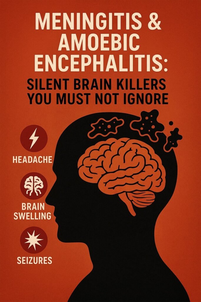
How Dangerous Is Meningitis?

Meningitis is still one of the most serious brain infections in the world.
• Even with treatment, the death rate can be as high as 25%.
• Survivors may face lifelong difficulties such as seizures, hearing loss, or paralysis.
 
A rarer and even deadlier form is Primary Amoebic Meningoencephalitis (PAM).

• It is caused mainly by Naegleria fowleri, and sometimes by Entamoeba histolytica.
• These organisms enter through the nose, travel via the olfactory nerve (responsible for smell), and destroy brain tissue.
• The risk is much higher: more than 90% of patients die, often within days, because the disease progresses so rapidly that doctors have very little time to act.
 
This is why meningitis and encephalitis are not merely “medical problems”; they are serious global health threats.

Warning Signs to Watch For

Most problems arise because infection causes cerebral edema (swelling of the brain) and raised intracranial pressure (ICP). This pressure damages brain cells and stops them from working normally.
 
🔴 Common symptoms include:
• Severe, sudden headache
• Vomiting, sometimes without nausea
• Sleepiness, confusion, or drowsiness from brain swelling
• Seizures (fits)
• Stiff neck, fever, or sensitivity to light
• In children: unusual crying, irritability, or refusal to feed
 
⚠️ If any of these symptoms appear, early diagnosis and immediate treatment are the key to survival. Delays can waste precious time and make the outcome worse.

Why Are We Hearing About Kerala?

Recently, there have been reports of amoebic encephalitis in Kerala.
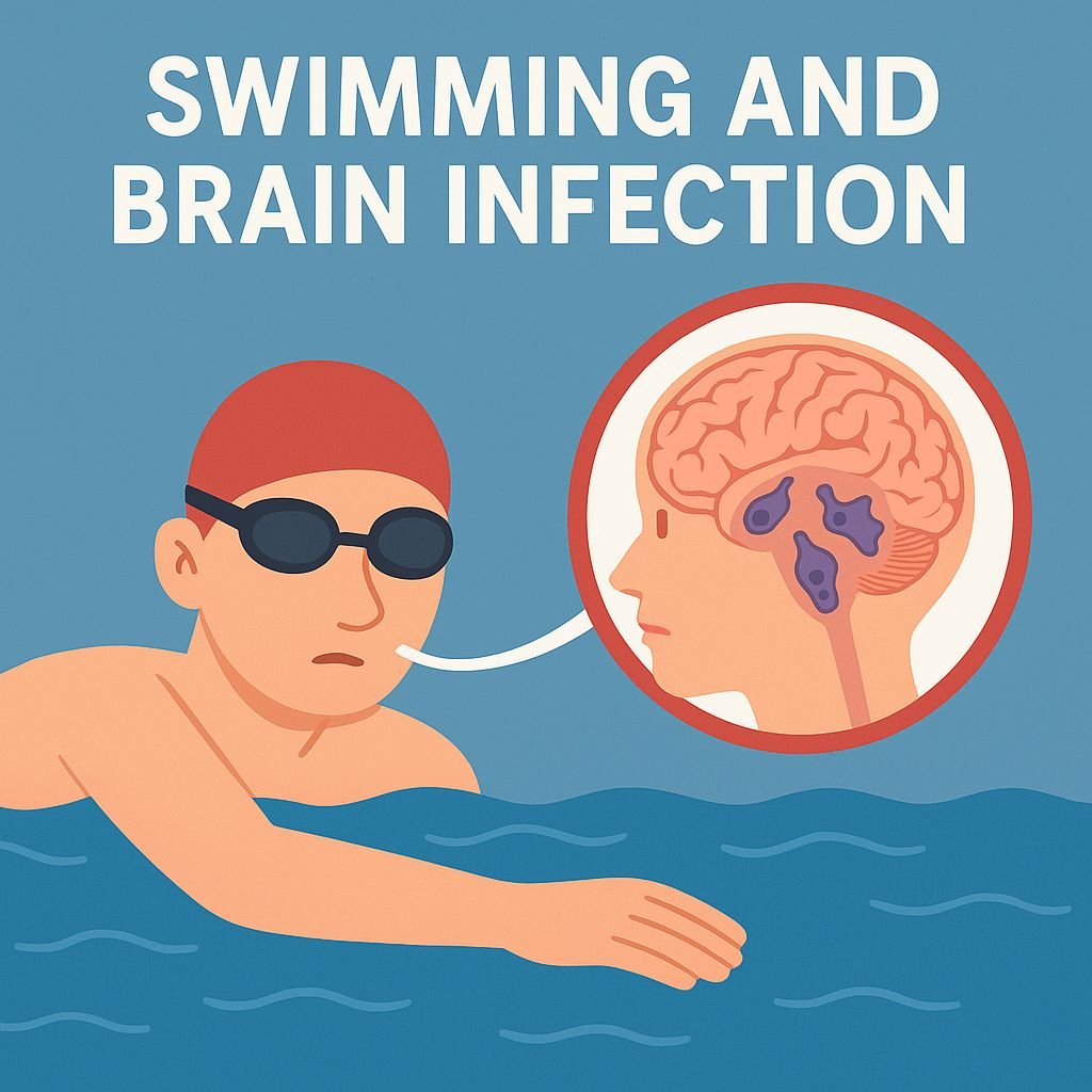
PAM is usually from exposure to unclean or poorly chlorinated water in ponds, lakes, and swimming pools.

When someone dives or swims in such water, the amoeba can enter through the nose. From there, it travels to the brain, causing severe swelling and inflammation. Sadly, most cases progress so fast that they are fatal within days of symptoms starting.

Who Is at Higher Risk?

• Children and elderly people, because of weaker immune systems.
• Patients with chronic illnesses or those on medicines that weaken immunity (such as steroids, cancer treatment, or after organ transplants).
• Anyone exposed to unsafe or untested water sources.

Dr. Ganesh Veerabhadraiah

Consultant – Neurosurgeon, Neurointerventional Surgery, Spine Surgeon (Neuro)
23+ Years Experience Overall (17+ years as Neuro Specialist)

Available for Consultation: Jayanagar 9th Block & Kauvery Hospital, Electronic City 

How Can We Protect Ourselves?

✅ Swim only in safe, chlorinated pools.
✅ Avoid diving into lakes or ponds with uncertain water quality.
✅ If swimming in unclear water, use nose clips — or better, avoid such exposure altogether.
✅ Shower after swimming, wash your hands, and clean clothes and towels properly.
✅ Drink only clean, purified water.
✅ Keep children, elderly, and immunocompromised people away from risky swimming environments.
 
The Take-Home Message
• 1 in 4 people with meningitis may die.
• 9 out of 10 people with amoebic encephalitis do not survive.
 
These are not just numbers. They represent real families losing loved ones.
 
The good news is that prevention and quick action save lives.
• Watch out for the signs.
• Seek medical care immediately.
• Choose safe water and healthy habits.
 
🧠 Your brain is priceless. Protect it — because a few simple choices could mean the difference between life and death.

Conclusion

Meningitis and amoebic encephalitis are not rare health issues — they are global threats. With mortality rates as high as 25% for meningitis and 90% for amoebic encephalitis, prevention is the best protection.

Choose safe water
Recognize early warning signs
Seek immediate medical care

Don’t wait until it’s too late. If you or a loved one shows symptoms of severe headache, seizures, or confusion,

FAQs

1) What are the first symptoms of meningitis?

Severe headache, vomiting, stiff neck, fever, confusion, and seizures are common warning signs.

2) How does amoebic encephalitis spread?

It spreads when contaminated water carrying Naegleria fowleri enters through the nose and travels to the brain.

3) Who is at higher risk of brain infections?

Children, elderly people, and those with weakened immunity (due to chronic illness, steroids, or cancer treatment) face higher risk.

4) Can meningitis and amoebic encephalitis be prevented?

Yes. Avoid unsafe water sources, swim only in chlorinated pools, and seek immediate medical help if symptoms appear.

5) What is the survival rate of amoebic encephalitis?

Unfortunately, more than 90% of patients do not survive, making prevention and rapid diagnosis essential.

 Surgical Management of a Colloid Cyst of third ventricle

Surgical Management of a Colloid Cyst of third ventricle

A 20-year-old girl, an art student, presented with two episodes of transient loss of consciousness lasting 20–30 minutes, accompanied by neck pain radiating to the left side. Clinical evaluation, followed by MRI, revealed a third ventricular colloid cyst, causing obstruction at the foramen of Monro and resulting in intermittent symptoms of raised intracranial pressure.

The patient underwent neuro-navigation-guided craniotomy and successful excision of the colloid cyst. Intraoperative and postoperative management were uneventful. The patient was extubated immediately following the procedure. A postoperative CT scan confirmed complete excision of the cyst, with no residual tumor visible.

She was monitored in the ICU for one day, where she demonstrated remarkable recovery. Impressively, she resumed her artistic pursuits on the first postoperative day, highlighting her swift neurological recovery. Currently, she is stable and recovering well in the ward.

We extend our gratitude to the entire neurosurgery team and the operating theater staff for their seamless coordination and dedication to achieving an excellent outcome for the patient.

This case underscores the importance of timely diagnosis, precise surgical planning, and multidisciplinary teamwork in the successful management of colloid cysts.

Histopathology- Nimhans 

Colloid cyst 

Regarding Third ventricular colloid cysts , these are rare 1 to 3 % whole intracranial tumours but potentially life-threatening intracranial lesions, often presenting a significant surgical challenge due to their deep midline location and proximity to critical neurovascular structures. These cysts can cause acute hydrocephalus and elevated intracranial pressure through obstruction of the foramen of Monro, leading to symptoms such as severe headaches, intermittent loss of consciousness, or, in rare cases, sudden death. Surgical management, either through neuro-navigation-guided craniotomy or endoscopic resection, demands meticulous planning and precise execution to minimize complications. Advances in neuro-navigation and microsurgical techniques have significantly enhanced the safety and efficacy of these procedures, making early diagnosis and prompt intervention essential in preventing catastrophic events.

Dr Ganesh Veerabhadraiah 
HOD and Senior Consultant Neurosurgeon
Kauvery hospital 
Electronic City
Bengaluru 

Ph no 7249669911

#ColloidCyst #ThirdVentricularTumours #BrainTumours #IntraventricularTumours #EndoscopicTumourRemoval #Craniotomy

TLIF Spinal Fusion Surgery: 9 Must-Know Tips Before You Decide

If you’ve been advised to undergo a TLIF (Transforaminal Lumbar Interbody Fusion) surgery, here are some steps you can take:

1. Understand the Procedure

ㆍLearn about what TLIF surgery entails, including the reasons it’s recommended, the benefits, and the risks involved.

2.Ask Questions

ㆍDiscuss any concerns with your surgeon. Questions might include:

 – What are the expected outcomes?
– What are the potential risks and complications?
– What can you expect in terms of recovery time?
– if surgery is open or  minimally invasive
 – if hospital is you are undergoing is infrastructured to undergo procedure and all necessary equipments are there.  

3. Prepare for Surgery

ㆍ Follow any pre-surgery instructions given by your doctor, such as:
ㆍ Avoiding certain medications (like blood thinners).
Dietary changes or fasting before the procedure.

4. Plan for Recovery

ㆍArrange for help at home after the surgery, as you may have restrictions on movement.
ㆍPrepare your living space for comfort and safety during recovery.

5. Follow Post-operative Care Instructions

ㆍAdhere to your doctor’s guidelines for post-surgery care, including physical therapy if recommended, pain management, and activity restrictions.

6.Stay Informed

Keep up with follow-up appointments and any recommended imaging or tests to monitor your recovery. 

7. Get a Second Opinion 

 If you feel uncertain, consider consulting another spine specialist to confirm the need for surgery and explore all treatment options. 

Insurance – usually all insurances are not covered by all hospitals- If your surgery is covered by insurence,  usually service line managers ( Neuro SLM ) will give information regarding this.  

8. Consider Lifestyle Changes

 Discuss long-term strategies for maintaining spine health with your healthcare provider, including exercise, weight management, and ergonomics in daily activities.

Always prioritize communication with your Neurosurgery team and make informed decisions that align with your health needs and goals.

Dr. Ganesh Veerabhadraiah

Consultant – Neurosurgeon, Neurointerventional Surgery, Spine Surgeon (Neuro)
23+ Years Experience Overall (17+ years as Neuro Specialist)

Available for Consultation: Jayanagar 9th Block & Kauvery Hospital, Electronic City 

FAQs

1. What is the success rate of TLIF surgery?

TLIF surgery has a success rate of around 80%–90% for relieving chronic lower back pain caused by degenerative disc disease or spinal instability.

2. Is TLIF surgery safe?

Yes, TLIF surgery is generally safe when performed by an experienced spine surgeon. Like all surgeries, it carries some risks, but major complications are rare.

3. How long is the recovery after TLIF surgery?

Most patients can return to light activities in 4–6 weeks. Full recovery may take up to 3–6 months depending on overall health and rehabilitation.

What is spondylolisthesis?

Lower back pain may be caused by spondylolisthesis, a spinal disorder. It happens when one of your vertebrae or spine bones slips out of place and lands on the vertebra below it. Nonsurgical treatment can usually alleviate your problems. In most cases, surgery for severe grade spondylolisthesis is effective.

What is spondylolisthesis?

Spondylolsthesis is a condition in which the vertebrae move more than they should, resulting in spine instability. A vertebra slips out of place and lands on the one below it. There will be pars interarticularis defect a bone connecting upper and lower facet. It could exert pressure on a nerve, resulting in lower back or leg pain.

The Greek words spondylos, which means “spine” or “vertebra” and listhesis, which means “slipping, sliding or moving” are combined to form spondylolisthesis (pronounced sphon-di-low-less-THEE-sis).

Types of spondylolisthesis:

  •  Degenerative spondylolisthesis: the most prevalent kind is degenerative spondylolisthesis, which occurs as a result of ageing.
  • Isthmic spondylolisthesis: as a result of spondylolisthesis, isthmic spondylolistheis develops. The bone is weakened by the crack or fracture.
  • Congenital spondylolisthesis: when a baby’s spine does not form properly before birth, this is known as congenital spondylolisthesis. The person’s vertebrae are misaligned, putting them at risk for slippage later in life.
  • Less common types of spondylolisthesis are:
  1. Traumatic spondylolisthesis: it occurs when the vertebrae slip as a result of an injury.
  2. Pathological spondylolisthesis: when a disease, such as osteoporosis, or a tumour, develops pathological spondylolisthesis.
  3. Post surgical spondylolisthesis: slippage as a result of spinal surgery is known as post-surgical spondylolisthesis.

How common is spondylolisthesis?

Spondylolisthesis and spondylolysis affect approximately 4% to 6% of the adult population. It is easy to go years without realising you have spondylolisthesis because you do not have any symptoms.

Degenerative spondylolisthesis (which happens as a result of ageing and wear and tear of the spine) is more common in women than in men after the age of 50.

Isthmic spondylolisthesis (typically induced by spondylolysis) is one of the most common reasons for back pain in teenagers.

Causes of spondylolisthesis:

One of the most common causes of spondylolisthesis in young athletes is overextending the spine. Genetics could also have a role. Some people are born with spinal bones that are thinner than others. Wear and tear on the spine and discs (the cushions between the vertebrae) can develop this problem in older persons.

What are the symptoms of spondylolisthesis?

You might not notice any signs or symptoms of spondylolisthesis. Some people are unaware that they have the disease. If you do experience symptoms, the most common one is lower back ache. The discomfort may spread to the buttocks and thighs. You may also experience:

  • Back pain/leg pain aggravated by activity- working at home or bending and picking up object.
  • Stiffness in the back
  • Spasms in the hamstring muscle (muscles in the back of the thighs)
  • Walking or standing for long periods of time is difficult
  • When leaning over, there is pain
  • Numbness, tingling or weakness in the foot.

How is spondylolisthesis diagnosed?

Your doctor will conduct a physical examination and inquire about your symptoms. An imaging/scan will very certainly be required to confirm the diagnosis.

What imaging tests will be needed?

  • A spinal X ray allows doctors to see if a vertebrae is misaligned.
  • To see the spine in greater detail or to see soft tissue like discs and nerves, a CT scan or an MRI scan may be required.

How to reduce the risk of spondylolisthesis?

You can lower your chance of spondylolisthesis by doing the following:

  • Regularly exercise your back and abdominal muscles to keep them strong
  • Maintain a healthy body mass index (BMI). Weight gain puts additional strain on your lower back
  • To keep your bones well nourished and robust, eat a well balanced diet.

What is the outlook for people with spondylolisthesis?

 The success rate of surgery is very high. Spondylolisthesis surgery patients typically return to an active lifestyle after a few months following surgery. Following surgery, you will almost certainly require therapy to help you regain full function.

Summary:

Spondylolisthesis is one of the  common cause of back pain /discomfort, however it is not life threatening. There are a variety of therapists available, ranging from medicine to physical therapy to spinal surgery. If you are experiencing low back discomfort or finding it difficult to walk, stand, or bend over, consult the Neuro Wellness Care Center, they will provide the best and cost friendly Back Pain Treatment in Bangalore.

    DOWNLOAD EBOOK

      DOWNLOAD EBOOK

        DOWNLOAD EBOOK

          DOWNLOAD EBOOK

            DOWNLOAD EBOOK

              DOWNLOAD EBOOK