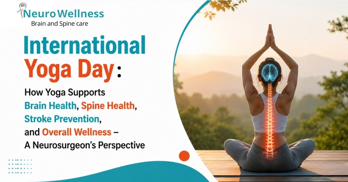
Every year, International Yoga Day reminds us of a timeless truth: our bodies are designed to move.
As a neurosurgeon, I spend my days treating conditions affecting the brain, spine, nerves, and blood vessels. I see patients suffering from neck pain, back pain, stroke, nerve compression, spinal disorders, poor posture, obesity-related complications, and lifestyle diseases. While modern medicine has made tremendous advances in treating these conditions, one question often comes to my mind:
Can some of these problems be prevented before they reach the operating room?
The answer, in many cases, is yes.
One of the simplest and most powerful tools available to us is regular physical activity, and yoga is one of the oldest and most effective forms of movement known to humanity.
Yoga: A Gift from Ancient Wisdom
Yoga is not a modern fitness trend.
It originated thousands of years ago in India and has been practiced by generations of sages, philosophers, and ordinary people seeking physical health, mental clarity, and spiritual well-being.
Long before modern medical science understood the importance of flexibility, breathing exercises, stress reduction, and movement, our ancestors had already recognized their value.
Today, scientific research continues to validate many of the benefits that yoga practitioners have experienced for centuries.
Yoga is more than a series of postures.
It is a holistic approach that combines:
* Physical movement
* Stretching
* Breathing techniques
* Mental focus
* Relaxation
* Self-awareness
Together, these elements create a powerful system for maintaining health and preventing disease.
Understanding Health at the Cellular Level
When most people think about health, they think about organs.
They think about the brain, heart, lungs, kidneys, liver, muscles, or joints.
However, every organ in the body is made up of millions and millions of individual cells.
The true foundation of health lies at the cellular level.
Every cell requires:
* Oxygen
* Nutrients
* Water
* Healthy blood circulation
* Proper waste removal
When these requirements are met, cells function efficiently.
Healthy cells create healthy tissues.
Healthy tissues create healthy organs.
Healthy organs create a healthy human being.
When circulation becomes poor, when movement decreases, or when metabolic disorders develop, cells begin to function less efficiently.
Over time, this can contribute to disease.
This is where yoga and regular physical activity become extremely important.
The Importance of Blood Flow
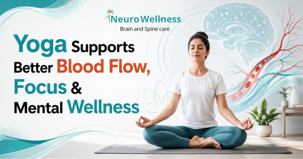
One of the most critical functions in the human body is circulation.
The heart pumps blood continuously throughout our lives.
Every heartbeat delivers oxygen and nutrients to every organ.
The brain, in particular, is highly dependent on uninterrupted blood flow.
Although the brain represents only a small percentage of body weight, it consumes a significant amount of the body’s oxygen and energy.
Even a brief interruption in blood flow can have serious consequences.
Stroke: A Powerful Example
As a neurosurgeon, I frequently treat patients suffering from stroke.
A stroke occurs when blood flow to a part of the brain becomes blocked or interrupted.
Without oxygen and nutrients, brain cells begin to die.
The consequences can include:
* Weakness of the arm or leg
* Difficulty speaking
* Facial drooping
* Memory problems
* Loss of independence
* Permanent disability
Many stroke risk factors are associated with lifestyle habits, including:
* High blood pressure
* Diabetes
* Obesity
* Physical inactivity
* Smoking
* High cholesterol
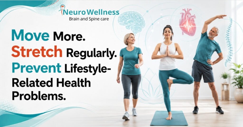
While yoga is not a guarantee against stroke, maintaining an active lifestyle can contribute significantly to better cardiovascular health and overall well-being.
The Modern Lifestyle Crisis
Human beings were never meant to sit for 8–12 hours every day.
Yet this has become the reality for millions of people.
Office workers, IT professionals, students, business owners, and even children spend prolonged periods sitting.
Modern technology has made life easier but often less active.
Many people spend their day:
* Sitting at a desk
* Looking at a computer screen
* Using a smartphone
* Watching television
* Travelling in vehicles
This sedentary lifestyle affects every system in the body.
Over time, it contributes to:
* Neck pain
* Back pain
* Obesity
* Poor posture
* Muscle weakness
* Diabetes
* Hypertension
* Heart disease
* Stress and anxiety
The body gradually loses flexibility, strength, and endurance.
The consequences may not appear immediately, but they accumulate over years.
Why the Spine Loves Movement
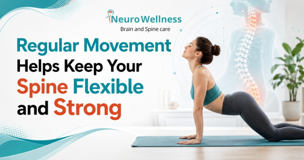
The spine is one of the most remarkable structures in the human body.
It supports the body, protects the spinal cord, and allows movement in multiple directions.
The spinal discs act as shock absorbers between vertebrae.
These discs depend on movement for nutrition.
Unlike many tissues, spinal discs do not have a direct blood supply.
They receive nutrients through a process that is enhanced by movement and changes in pressure.
Prolonged sitting can contribute to:
* Muscle stiffness
* Disc degeneration
* Poor posture
* Neck pain
* Lower back pain
Gentle stretching and regular movement help maintain flexibility and support spinal health.
This is one reason why many people experience improvement in stiffness and discomfort after adopting a regular yoga routine.
Yoga and Posture
Poor posture has become one of the most common health issues in modern society.
Forward head posture, rounded shoulders, and prolonged sitting place additional stress on the neck and spine.
Many individuals develop:
* Chronic neck pain
* Shoulder pain
* Upper back discomfort
* Headaches
* Muscle fatigue
Yoga encourages awareness of body position.
Through stretching and strengthening exercises, it helps improve posture and balance.
Better posture reduces unnecessary stress on muscles and joints.
Yoga and Mental Health
Health is not only physical.
The brain is affected by stress just as much as the body.
Modern life exposes us to constant pressure:
* Work deadlines
* Financial concerns
* Family responsibilities
* Social expectations
* Digital overload
Chronic stress can contribute to:
* Anxiety
* Poor sleep
* Fatigue
* Reduced concentration
* Mood disturbances
Breathing exercises and mindfulness practices associated with yoga may help calm the nervous system.
Many individuals report improved:
* Focus
* Emotional balance
* Sleep quality
* Mental clarity
As a brain specialist, I believe mental wellness is just as important as physical wellness.
A healthy brain supports a healthy life.
Breathing: The Forgotten Medicine
Most people rarely think about their breathing.
Yet breathing is fundamental to life.
Every cell depends on oxygen.
Yoga emphasizes conscious breathing techniques.
Deep breathing encourages:
* Better oxygen delivery
* Relaxation
* Reduced stress response
* Improved awareness
When combined with physical movement, breathing exercises become a powerful tool for overall wellness.
Yoga and the Aging Process
As we age, the body naturally undergoes changes.
Flexibility decreases.
Muscle mass declines.
Balance may become less stable.
Joint stiffness becomes more common.
Many people mistakenly assume these changes are unavoidable.
While aging cannot be stopped, healthy habits can help maintain function and independence.
Regular movement helps preserve:
* Strength
* Balance
* Flexibility
* Coordination
* Confidence
Many older adults find yoga particularly beneficial because it can be adapted to different fitness levels.
Can Yoga Prevent Disease?
Yoga is not a replacement for medical treatment.
Nor should it be viewed as a cure for all illnesses.
However, when practiced safely and consistently, yoga can be an important component of a healthy lifestyle.
Combined with:
* Good nutrition
* Adequate sleep
* Hydration
* Stress management
* Medical care when needed
Yoga may contribute to reducing the risk of many lifestyle-related conditions.
The goal is not perfection.
The goal is prevention.
Movement is Medicine
One of the most important messages I share with patients is simple:
The body needs movement.
It does not necessarily have to be intense.
You do not need to become an athlete.
You do not need expensive equipment.
You simply need consistency.
Whether it is:
* Yoga
* Walking
* Swimming
* Cycling
* Stretching
* Strength training
Regular physical activity supports long-term health.
The best exercise is often the one you can continue doing consistently.
What About People with Medical Conditions?
Many patients ask whether yoga is safe for them.
The answer depends on the individual condition.
Patients with:
* Severe spinal disorders
* Recent surgery
* Significant neurological conditions
* Severe arthritis
* Balance problems
Should consult their healthcare provider before starting any exercise program.
Not every posture is suitable for every individual.
Yoga should be adapted according to age, flexibility, fitness level, and medical condition.
Safety should always come first.
A Message to Young People
One of the greatest concerns today is the declining level of physical activity among younger generations.
Children and teenagers spend increasing amounts of time:
* On smartphones
* On tablets
* Playing video games
* Sitting indoors
Healthy habits formed during childhood often continue into adulthood.
Encouraging physical activity early in life can have lifelong benefits.
Yoga can help young people develop:
* Discipline
* Flexibility
* Concentration
* Body awareness
* Stress management skills
These benefits extend far beyond physical fitness.
A Message to Corporate Employees
Many IT professionals and office workers spend 8–12 hours sitting each day.
This places tremendous strain on the neck, back, shoulders, and eyes.
Small daily habits can make a significant difference:
* Stretch every hour
* Take walking breaks
* Maintain good posture
* Practice breathing exercises
* Incorporate yoga into your weekly routine
Your future health depends on the choices you make today.
International Yoga Day: A Call to Action
International Yoga Day is not just about performing a few postures for one day.
It is about embracing a lifestyle that values movement, balance, and self-care.
The goal is not to achieve difficult poses.
The goal is to take care of the body and mind that carry us through life.
Every stretch matters.
Every step matters.
Every healthy choice matters.
Final Thoughts from a Neurosurgeon
As a neurosurgeon, I have witnessed both the incredible resilience of the human body and the devastating consequences of neglecting health.
Modern medicine can treat many conditions.
Surgeons can perform complex procedures.
Hospitals can provide advanced care.
But prevention remains the most powerful medicine.
Yoga reminds us of a simple truth that has existed for thousands of years:
A healthy mind resides in a healthy body.
By staying active, stretching regularly, breathing deeply, and caring for ourselves at the cellular level, we invest in our future health.
On this International Yoga Day, let us renew our commitment to movement, wellness, and prevention.
Because the goal is not merely to live longer.
The goal is to live healthier, stronger, and more meaningful lives.
Move more. Stretch more. Breathe better. Live healthier.
Happy International Yoga Day.
– Dr. Ganesh Veerabhadraiah
Senior Consultant Neurosurgeon & Head of Department – Neurosurgery
Founder, NeuroWellness
Brain • Spine • Stroke Care
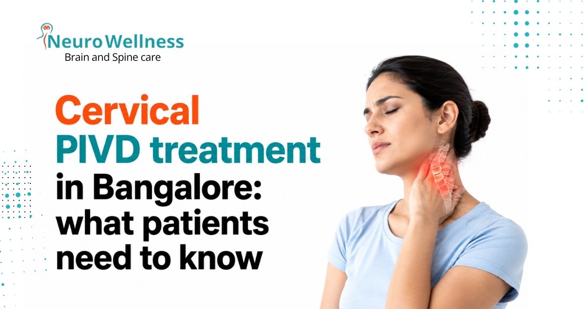
Questions about your cervical collar after surgery?
Dr Ganesh Veerabhadraiah and Dr Sharan Srinivasan at NeuroWellness Brain and Spine Clinic, Jayanagar, provide post-operative follow-up care for all cervical spine surgeries. If you have concerns about your collar, your recovery timeline, or your follow-up imaging, our team is available for consultations.
G-Floor, 26th Main, 9th Block Jayanagar, Bengaluru 560069 | Monday–Saturday
Related reading from NeuroWellness Brain and Spine Clinic
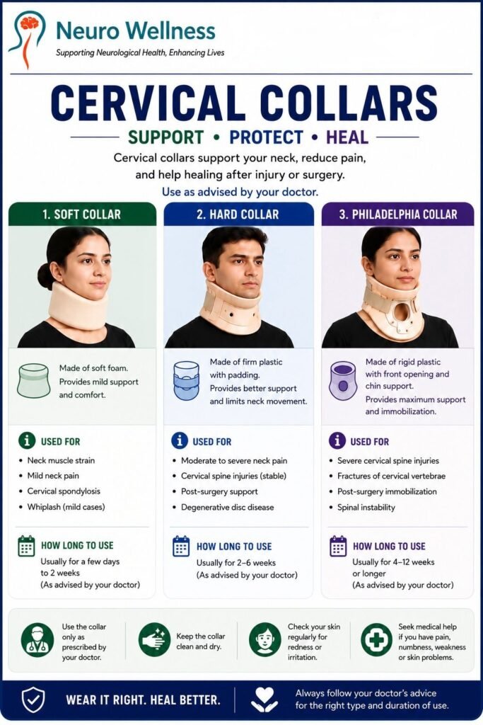
Cervical collar types: soft, hard and Philadelphia — complete guide
Neck collar for cervical spondylosis: which type, how long and what to avoid
Cervical PIVD treatment in Bangalore: symptoms, non-surgical and surgical options
Exercises to relieve neck pain: 12 safe moves for after collar removal
Dr Ganesh Veerabhadraiah — Senior Neurosurgeon, NeuroWellness Jayanagar