Cervical PIVD a prolapsed disc in the neck is one of the most common reasons patients visit a neurosurgeon in Bangalore. At NeuroWellness Brain and Spine Clinic, Jayanagar, we see patients every week who have been living with arm pain, hand tingling, or neck stiffness caused by cervical disc compression, often without knowing that effective treatment surgical and non-surgical is available close to home. This guide explains what cervical PIVD is, how it is diagnosed, what treatment involves, and when to act.
What is cervical PIVD?
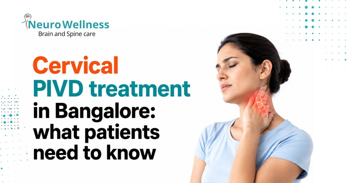
PIVD stands for Prolapsed Intervertebral Disc. Cervical PIVD is when a disc in the neck ruptures and its inner nucleus pushes outward, compressing a nearby nerve root or the spinal cord. It causes neck pain, arm pain, hand numbness, and in severe cases weakness or gait problems. It most commonly affects the C5–C6 and C6–C7 disc levels.
Each vertebra in the cervical spine is separated by an intervertebral disc a shock-absorbing structure with a tough outer ring (annulus fibrosus) and a soft gel-like centre (nucleus pulposus). When the disc degenerates or is stressed — due to age, injury, or sustained poor posture — the outer ring can develop cracks or tears through which the inner nucleus pushes outward. This is the prolapse.
When the prolapsed disc material presses on a nerve root exiting the spine, it causes pain, tingling, and weakness in the arm — a pattern called cervical radiculopathy. When it presses on the spinal cord itself, it can affect walking, balance, and fine motor function a more serious condition called cervical myelopathy that requires prompt neurosurgical assessment.
C5–C6
Most commonly affected disc level in cervical PIVD
80%
of cervical PIVD cases resolve without surgery with appropriate conservative care
6–12 wks
typical timeline for conservative treatment trial before surgical reassessment
Cervical PIVD vs lumbar PIVD - key difference
Lumbar PIVD (lower back disc prolapse) is more common overall, but cervical PIVD carries a higher risk of serious neurological complication. A prolapsed lumbar disc compresses nerve roots serving the legs. A prolapsed cervical disc compresses nerve roots serving the arms and can also compress the spinal cord, which carries signals for the entire body below the neck. This is why cervical PIVD with cord involvement is treated more urgently than most lumbar disc problems.
Which cervical levels are most commonly affected
Cervical PIVD symptoms - what patients report
The symptoms of cervical PIVD vary depending on which disc level is affected and whether nerve roots or the spinal cord is involved. Most patients present with a combination of neck symptoms and symptoms in one or both arms.
Neck pain and stiffness
Typically the earliest symptom a dull or aching pain at the back of the neck, often worse with certain movements, prolonged sitting, or screen use. Neck stiffness, particularly in the morning, is common. The pain may worsen suddenly if the disc prolapse is acute or traumatic.
Arm pain (cervical radiculopathy)
A sharp, shooting, burning, or electric pain that travels from the neck down the arm — the specific path depends on which nerve root is compressed. Many patients describe it as a "hot wire" sensation running from the neck to the fingertips. This is the hallmark symptom of nerve root involvement.
Hand numbness and tingling
Numbness, pins and needles, or a "dead" feeling in specific fingers correlates to the affected nerve root level. The distribution is distinctive — index finger numbness points to C6, middle finger to C7, and little finger to C8. Patients often describe waking at night with a numb hand.
Weakness in grip or shoulder
Reduced grip strength, difficulty opening jars, dropping objects, or weakness when raising the arm — these reflect motor nerve root compression. Weakness is a more serious symptom than pain or numbness alone and warrants earlier specialist review. Wasting of hand muscles indicates prolonged compression.
Myelopathy symptoms - when the spinal cord is compressed
- Difficulty walking or unsteady gait — patients describe feeling drunk or as if the legs “don’t quite work” on uneven surfaces
- Difficulty with fine motor tasks — inability to button a shirt, write legibly, or type with normal speed
- Weakness in both arms or both legs — bilateral symptoms always raise concern for spinal cord involvement
- Lhermitte’s sign — an electric shock sensation running down the spine and into the limbs when the chin is dropped to the chest
- Bladder or bowel changes — urgency, difficulty starting urination, or incontinence alongside neck symptoms is a neurosurgical emergency
Cervical myelopathy — spinal cord compression from cervical disc disease — can worsen progressively and silently. Unlike radiculopathy, which often causes severe pain that drives patients to seek care early, myelopathy can progress gradually with symptoms patients initially attribute to ageing. Once spinal cord damage becomes permanent, it is irreversible. Early surgical decompression is the only treatment.
How is cervical PIVD diagnosed?
Diagnosis of cervical PIVD involves a combination of clinical examination, imaging, and — in some cases — nerve function testing. The clinical examination tells the neurosurgeon which nerve root is likely involved; the imaging confirms the disc level and severity of compression.
Clinical neurological examination First step
Your neurosurgeon assesses reflexes, muscle strength in specific muscle groups, and the distribution of numbness across the arm and hand. Provocation tests — such as Spurling's test, where the neck is tilted toward the affected side under gentle pressure — can reproduce symptoms and identify the likely nerve root involved. The clinical exam often localises the problem before any imaging is ordered.
MRI cervical spine Gold standard
MRI is the definitive investigation for cervical PIVD. It shows the prolapsed disc, the degree of nerve root or spinal cord compression, the levels involved, and whether there are additional factors such as spondylotic narrowing or ligament thickening contributing to the compression. An MRI of the cervical spine is typically the first imaging investigation ordered when cervical PIVD is clinically suspected. Most major hospitals in Bangalore offer same-day or next-day MRI.
EMG and nerve conduction velocity (NCV) Selected cases
Electromyography (EMG) and nerve conduction velocity testing measure the electrical activity of muscles and the speed of nerve signals. They are useful when the clinical picture and MRI findings don't fully align, when there is a question of whether symptoms come from the cervical spine or from a peripheral nerve problem such as carpal tunnel syndrome, or when the duration and severity of nerve damage needs objective quantification before surgery.
X-ray cervical spine Limited role
Plain X-rays of the cervical spine show bony structures — vertebral alignment, disc space height, and osteophytes — but cannot show the disc material or nerve compression directly. X-rays are useful for assessing spinal alignment, ruling out fracture, and for pre-operative planning to assess disc space height and alignment. They cannot diagnose PIVD and should not be the only investigation when neurological symptoms are present.
Non-surgical treatment for cervical PIVD
The majority of cervical PIVD cases — approximately 75–80% — can be managed successfully without surgery when there is no spinal cord compression and neurological deficits are not progressive. Non-surgical treatment is the appropriate first step for most patients and involves a structured combination of the following measures.
Rest and activity modification
During the acute phase of cervical PIVD — typically the first 1 to 2 weeks — reducing activities that aggravate symptoms allows the inflammatory response around the compressed nerve to begin settling. This does not mean complete bed rest, which is counterproductive. It means avoiding activities that provoke arm pain: overhead reaching, heavy lifting, sustained screen use with poor posture, or driving for long periods. A brief period of activity modification combined with pain relief gives the disc the chance to partially retract and the nerve the opportunity to begin recovery. Duration: 1–2 weeks acute rest, then graduated return to activity under physiotherapy guidance.
Cervical collar use
A soft cervical collar is often prescribed during the first 2 to 4 weeks of acute cervical PIVD management. It reduces neck movements that reproduce or worsen arm pain, provides mild support for the paraspinal muscles, and allows inflamed nerve roots brief respite from compressive movement. The collar is a short-term symptomatic measure — it does not treat the disc prolapse and should not be worn beyond the prescribed period. For a complete guide to cervical collar use, see our cervical collar types guide and the neck collar for spondylosis patient guide. Duration: 2–4 weeks soft collar. Do not self-extend beyond prescription.
Physiotherapy and cervical traction
Physiotherapy for cervical PIVD focuses on two goals: reducing nerve compression and strengthening the muscles that support the cervical spine. Manual therapy, gentle mobilisation, and — when appropriate — mechanical or manual cervical traction are used. Traction creates slight distraction between vertebrae, temporarily reducing the pressure on the prolapsed disc and providing relief for many patients with radiculopathy. Specific exercises — particularly isometric neck strengthening — are introduced once acute pain settles. See our neck pain exercise guide for the exercises most appropriate for cervical conditions. Begin under physiotherapist supervision — some exercises worsen disc compression if performed incorrectly.
Medications
NSAIDs (anti-inflammatory drugs): reduce inflammation around the compressed nerve root and provide pain relief. First-line pharmacological treatment. Stomach protection (PPI) is typically co-prescribed. Muscle relaxants: reduce the muscle spasm that often accompanies cervical PIVD and contributes to pain. Short-term use only — typically 1 to 2 weeks. Neuropathic pain agents: Pregabalin or Gabapentin are often prescribed for the burning, shooting, or electric arm pain of cervical radiculopathy. They target the nerve pain specifically and are more effective for this symptom type than standard NSAIDs alone. Oral corticosteroids: a short reducing course of oral steroids (such as methylprednisolone) can provide rapid relief for severe acute radiculopathy by reducing the perineural inflammation dramatically. Used selectively in severe presentations. All medication must be prescribed by a qualified doctor — do not self-medicate for cervical PIVD symptoms.
Cervical epidural steroid injection
A cervical epidural steroid injection (CESI) delivers corticosteroid medication directly into the epidural space around the compressed nerve root under fluoroscopic (X-ray) or CT guidance. It provides targeted anti-inflammatory effect at the exact site of nerve compression, often producing significant and rapid relief of radicular arm pain. CESI is used when oral medication and physiotherapy have provided insufficient relief, and when the patient wishes to defer or avoid surgery. The procedure is performed as a day case and most patients notice improvement within 1 to 2 weeks of the injection. Effects can last several months, during which time physiotherapy can be progressed more effectively. Performed by experienced interventional neurosurgeons. Available at NeuroWellness — ask your consultant about suitability.
How long to try conservative treatment before considering surgery
If arm or hand weakness is present from the start — or develops or worsens during conservative treatment — do not wait the full 6 to 12 weeks. Progressive motor deficit is an indication for earlier surgical intervention. The window for full neurological recovery narrows with the duration of compression.
Surgical treatment for cervical PIVD in Bangalore
When is surgery recommended for cervical PIVD?
| CLINICAL SITUATION | SURGERY INDICATED? | URGENCY |
|---|---|---|
| Pain only, no neurological deficit, not responding to 8–12 weeks conservative treatment | Yes — consider | Elective |
| Radiculopathy with significant arm pain not controlled by medication or CESI | Yes — earlier consideration | Elective to semi-urgent |
| Progressive arm or hand weakness | Yes — earlier surgery | Semi-urgent (weeks) |
| Cervical myelopathy (spinal cord compression) — gait difficulty, hand clumsiness | Yes — do not delay | Urgent (days to weeks) |
| Bladder or bowel dysfunction from cervical cord compression | Yes — emergency | Emergency |
| Pain only, responding well to conservative treatment | No — continue conservative | Continue current plan |
ACDF - Anterior Cervical Discectomy and Fusion
ACDF is the most commonly performed surgical procedure for cervical PIVD in Bangalore and globally. The surgery is performed through a small incision at the front of the neck. The damaged disc is completely removed, the nerve root and/or spinal cord is decompressed, and the disc space is filled with a bone graft or implant. A titanium plate and screws are then fixed to the adjacent vertebrae to stabilise the segment while the bone heals and fuses.
The anterior approach — through the front of the neck — allows excellent visualisation of the disc and avoids disruption of the posterior neck muscles. The skin incision is typically 3–4 cm and is placed in a natural neck skin crease, healing with a near-invisible scar in most patients.
For single-level ACDF, the operation typically takes 60–90 minutes under general anaesthesia. For two or three-level procedures, 90–150 minutes. Most patients are able to sit up and walk on the day of or the day after surgery.
Cervical disc replacement (arthroplasty)
Cervical disc replacement (CDR) — also called cervical arthroplasty — removes the prolapsed disc through the same anterior approach as ACDF but replaces it with an artificial disc implant rather than fusing the vertebrae. The artificial disc allows continued movement at the operated level, which theoretically reduces the rate of adjacent segment degeneration over time compared to fusion.
CDR is most appropriate for younger patients (typically under 55), single-level disease, disc prolapse without significant spondylosis or facet joint arthritis at the operated level, and patients who wish to preserve cervical motion for occupational or lifestyle reasons. It is not suitable for all patients — your neurosurgeon will assess whether CDR or ACDF is more appropriate based on your imaging and clinical profile.
Recovery timelines are similar to ACDF. Unlike ACDF, there is no post-operative hard collar required in most CDR patients, as there is no fusion to protect.
Posterior cervical foraminotomy
Posterior cervical foraminotomy approaches the disc from the back of the neck. A small portion of the facet joint and bone at the affected level is removed to widen the foramen (the opening through which the nerve exits), relieving the nerve root compression. The disc itself is not removed — instead, the bone narrowing the nerve’s exit point is addressed.
This approach preserves motion at the operated segment (no fusion) and avoids the anterior approach. However, it is only appropriate for soft disc herniations that are compressing the nerve root laterally — not for central disc prolapses compressing the spinal cord, and not for disc herniations combined with significant spondylosis or central canal narrowing.
When selected for the right patient and disc level, posterior foraminotomy offers comparable neurological outcomes to ACDF with a shorter recovery and no fusion-related restrictions.
Cervical PIVD — particularly when it involves the spinal cord or multiple nerve roots — is best managed by a neurosurgeon with specific cervical spine training. Neurosurgeons train extensively in the anatomy and surgical management of neural structures. For cervical PIVD with cord compression (myelopathy), neurological expertise in the peri-operative period is essential. Both neurosurgeons and orthopaedic spine surgeons perform ACDF, but for complex cervical PIVD with neurological involvement, a neurosurgical assessment ensures the most comprehensive evaluation of your spinal cord and nerve root function.
Cervical PIVD treatment at NeuroWellness, Jayanagar, Bangalore
WHY PATIENTS ACROSS BANGALORE CHOOSE NEUROWELLNESS FOR CERVICAL PIVD
- Dedicated neurosurgical assessment — not a general orthopaedic clinic
- On-site MRI review and same-day consultation available
- Both conservative and surgical management under one team
- ACDF and cervical disc replacement performed by experienced spine neurosurgeons
- Serving patients from Jayanagar, JP Nagar, BTM, Banashankari, Bilekahalli and South Bangalore
- Transparent consultation — surgery is recommended only when clinically necessary
Jayanagar 9th Block, Bengaluru 560069 | Appointments Monday–Saturday
Frequently asked questions - cervical PIVD
1. What does PIVD mean in medical terms?
PIVD stands for Prolapsed Intervertebral Disc. It refers to a condition where the soft inner nucleus pulposus of a spinal disc pushes through the tough outer fibrous ring (annulus fibrosus), compressing nearby nerve roots or the spinal cord. When this occurs in the neck it is called cervical PIVD. It is also referred to as cervical disc herniation or cervical disc prolapse. The full form of PIVD in medical terminology is Prolapsed Intervertebral Disc.
2. Can cervical PIVD be cured without surgery?
Yes, the majority of cervical PIVD cases resolve with conservative treatment over 6 to 12 weeks. This includes rest and activity modification, cervical collar use during the acute phase, physiotherapy and cervical traction, anti-inflammatory and neuropathic pain medication, and in some cases cervical epidural steroid injections. Surgery is only considered when conservative treatment fails after an adequate trial, or when there is spinal cord compression (myelopathy) causing progressive weakness or gait problems — which requires earlier intervention.
3. How long does recovery from cervical PIVD surgery take?
Most patients undergoing ACDF surgery for cervical PIVD in Bangalore return to desk or office work within 4 to 6 weeks and resume full physical activity within 3 months. The cervical bone fusion takes approximately 3 to 6 months to consolidate on follow-up imaging, though most patients experience significant pain and neurological improvement well before this. Recovery varies based on the number of spinal levels operated and the duration and severity of pre-operative nerve compression — patients with longer-standing weakness may have slower neurological recovery.
4. Which doctor should I see for cervical PIVD in Bangalore?
A neurosurgeon or spinal neurosurgeon is the most appropriate specialist for cervical PIVD, particularly if you have arm pain, hand numbness, or arm weakness. At NeuroWellness Brain and Spine Clinic in Jayanagar, Bangalore, Dr Ganesh Veerabhadraiah and Dr Sharan Srinivasan specialise in cervical spine conditions including cervical disc prolapse, ACDF surgery, and cervical disc replacement. The clinic is located at G-Floor, 26th Main, 9th Block Jayanagar, Bengaluru 560069 and is accessible from JP Nagar, BTM Layout, Banashankari, and South Bangalore.
5. Is a cervical collar necessary for PIVD?
A soft cervical collar is often prescribed during the acute phase of cervical PIVD to reduce pain and limit neck movements that aggravate nerve compression. It is a temporary measure typically used for 2 to 4 weeks — it provides symptomatic relief while the disc and surrounding inflammation settle, but it does not treat the prolapse itself. A collar should not be worn indefinitely for PIVD. If you have had cervical surgery for PIVD, a hard collar will be prescribed for 6 to 12 weeks post-operatively depending on the procedure.
6. What happens if cervical PIVD is left untreated?
Untreated cervical PIVD with significant nerve root compression can progress to permanent nerve damage, persistent arm weakness, and chronic pain that becomes increasingly difficult to manage. In cases where the disc is compressing the spinal cord (cervical myelopathy), delayed treatment risks progressive and potentially irreversible loss of hand function, balance, and walking ability. Many cervical PIVD cases do improve spontaneously over months — but any PIVD causing neurological symptoms (arm weakness, numbness, gait difficulty) should be assessed by a neurosurgeon promptly rather than left without monitoring.
RELATED READING FROM NEUROWELLNESS BRAIN AND SPINE CLINIC
- Understanding disc prolapse at the neck — cervical PIVD explained
- Cervical collar types: soft, hard and Philadelphia — complete guide
- Neck collar for cervical spondylosis: which type, how long and what to avoid
- Exercises to relieve neck pain: 12 safe moves for home
- Dr Ganesh Veerabhadraiah — Senior Neurosurgeon, NeuroWellness Jayanagar Bangalore
Written by Dr Ganesh Veerabhadraiah Senior Neurosurgeon & Spine Specialist — NeuroWellness Brain and Spine Clinic, Jayanagar, Bangalore Published: June 2026