Strategies for Brain Stroke Survivors​

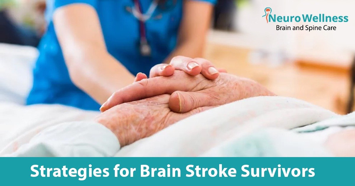
The human brain is a remarkable organ, controlling our thoughts, emotions, and movements. Yet, when struck by a stroke, this intricate network can be significantly disrupted, leaving survivors with various challenges to overcome. Every year, millions of individuals around the world face the life-altering effects of a brain stroke, leading to physical, cognitive, and emotional changes that require careful attention and rehabilitation. Life can change in an instant. Imagine going about your day when suddenly, a powerful force disrupts the intricate workings of your brain. A stroke, like a thief in the night, robs you of control, leaving behind a trail of physical and cognitive challenges. It’s an experience that many stroke survivors know all too well.
Stroke is the second most common cause of death in India, as per expert. About 5.2 million strokes were witnessed in children, aged less than 20 years. Stroke is the second most common cause of mortality in India, According to Dr. MV Padma Srivastava, Professor of Neurology at AIIMS. India reports over 1,85,000 incidents of stroke annually, with one stroke mortality occurring every four minutes and one stroke occurring every 40 seconds.

Understanding Brain Strokes:

A brain stroke occurs when the blood supply to the brain is interrupted, resulting in the rapid loss of brain function. This disruption can be caused by a blockage (ischemic stroke) or bleeding (hemorrhagic stroke) within the brain. Survivors often encounter physical, cognitive, and emotional challenges that necessitate comprehensive support and rehabilitation.

Strategies for Brain Stroke Survivors:

Let’s explore it more in detail;

Rehabilitation Therapy:

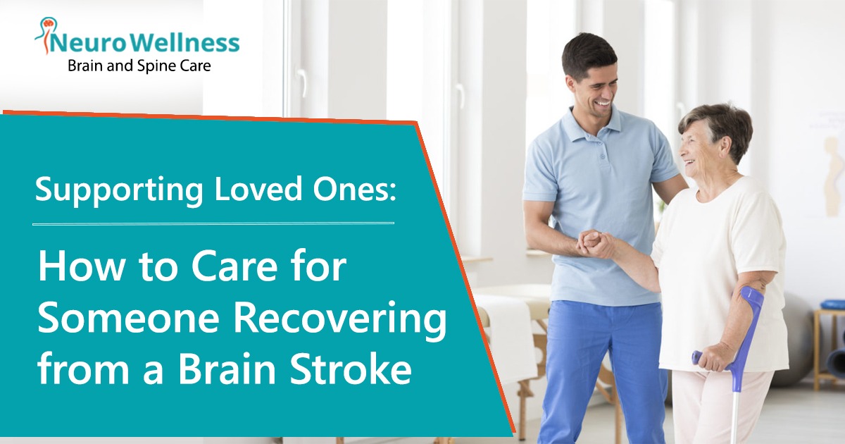
Rehabilitation therapy plays a crucial role in the recovery process. Physical therapy focuses on regaining strength, mobility, and coordination, helping survivors improve their physical abilities. Occupational therapy assists in relearning daily living skills and adapting to any physical limitations. Speech therapy can help with communication difficulties and swallowing problems. Working with a skilled team of therapists, survivors can make remarkable progress and regain their independence.

Healthy Lifestyle Choices:

Adopting a healthy lifestyle is key to optimizing recovery. Regular exercise, under the guidance of healthcare professionals, can improve cardiovascular health, strength, and overall well-being. Eating a balanced diet, rich in fruits, vegetables, whole grains, and lean proteins, provides essential nutrients for healing and supports brain health. Adequate rest and quality sleep are also important for recovery and rejuvenation.

Assistive Technologies:

A brain stroke survivor’s quality of life can be greatly enhanced by embracing assistive technologies. Mobility aids, communication tools, and gadgets that may be adjusted to meet different demands are some of these technologies. With the use of assistive technology, survivors can overcome their physical and verbal limitations, fostering independence and improving general wellbeing.

Lifestyle Modifications:

Making necessary modifications to the living environment can create a safer and more accessible space for brain stroke survivors. Simple adjustments such as installing handrails, grab bars, can prevent falls and enhance mobility. Organizing the living space to reduce clutter and improve navigation can also contribute to a sense of security and independence.

Medication Management:

It is essential to follow your prescribed medication regimen diligently. Medications can help manage underlying conditions, prevent future strokes, and control symptoms. Regularly consult with your healthcare provider to ensure you are taking the right medications and understand their potential side effects.

Social engagements:

Maintaining social connections and engaging in meaningful activities is vital for the well-being of brain stroke survivors. Participate in support groups, community programs, and social activities that align with personal interests and abilities. Connecting with others who have shared experiences can provide emotional support, encouragement, and a sense of belonging.

Mindfulness and Stress Reduction:

Practicing mindfulness techniques, such as deep breathing exercises, meditation, and relaxation techniques, can help reduce stress and promote overall well-being. Stress management is crucial, as excessive stress can hinder the recovery process. Consider joining mindfulness programs or engaging in activities that bring you joy and relaxation.

Conclusion:

As you navigate the path to recovery after a brain stroke, Neuro Wellness Brain and Spine Clinic is here to support you every step of the way. By implementing strategies such as rehabilitation therapy, Healthy Lifestyle Choices, Assistive Technologies Lifestyle Modifications, Medication Management, Social engagements, Mindfulness and Stress Reduction, you can unlock your potential for healing and regain independence. Remember, each survivor’s journey is unique, and at Neuro Wellness, we are dedicated to providing personalized care and guidance tailored to your specific needs. Let us embark on this transformative journey together, where hope thrives, and you rediscover the joy of life after a brain stroke.

Brain Stroke: Faster you act, the better you save the patient life

Dr Ganesh Veerabhadraiah

MBBS, DNB (Neurosurgery)

FINR (Fellowship in Interventional Neurosurgery-Switzerland)

As we know about Brain Stroke: Faster you act, the better you save the patient life. National, 17th April 2022: A brain stroke, also known as a brain attack, occurs when the supply of blood to the brain is reduced or stopped. It is like a heart attack. However, in a brain stroke, due to blockage inside the vessel, blood and oxygen supply to the brain are halted. A few of the common symptoms of a brain stroke are facial weakness, arm and leg weakness, speech dysfunction, blurring of vision, and imbalance while walking. For brain stroke Dr. Ganesh Veerabhadraiah is one of the top neurosurgeon in Bangalore.

What risk factors make the brain prone to stroke?

If you are suffering from hypertension, diabetes, smoking, high cholesterol, obesity, or ischemic heart disease, you are predisposed to a brain stroke. Today’s sedentary lifestyle is one of the major causes of the development of the above-mentioned diseases, so it is important to make changes in your lifestyle so that you can keep your body healthy and reduce the risk of developing these diseases.

Do we have a golden hour in a brain stroke as we have in a heart attack?

A brain stroke is very similar to a heart attack. Suppose a person suddenly develops chest pain, the first thing that comes to your mind is to take him or her to the hospital. As first aid, the medical team first got the ECG and ECHO done, and the cardiologist advised further treatment.

Similarly, in a brain attack or a brain stroke, complications can be avoided if the warning signs are recognized early and the person reaches the hospital within the GOLDEN HOUR.

Understand how a brain stroke happens:

So, as we know, the heart pumps blood into the brain through blood vessels called carotid arteries, which include the right and left carotids. If there is a blockage on one side of the brain and the blood supply is hindered, it may cause a brain stroke.

Studies show that during a brain stroke, 1.9 million neurons die every minute. This happens due to a lack of blood supply and if not treated immediately it might cause permanent damage to the important parts of the brain.

A brain stroke is further classified into two categories: mini-strokes and major strokes, based on the severity and the impact it causes on the brain.

If a person suffers mini strokes, his or her condition usually improves within 24 hours, while if a person suffers from a major stroke, he or she may take longer to recover. A major stroke might also result in weakness or neurological disorders for a longer time and sometimes permanent disability. To give you entire details and the best treatment Dr. Ganesh is one of the best Neurosurgeon in Bangalore with many years of experience.

What is this golden hour of Brain Stroke: Faster you act, the better you save the patient life?

When a brain stroke happens, it is imperative to take the patient to the hospital within the golden period. The golden hour is 3.5 hours to 7.2 hours after the person has suffered a brain stroke. If the clot-bursting medication and mechanical thrombectomy happen within this period, blood circulation re-establishes. Once blood circulation is restored, the supply of blood and nutrition will again resume, which will eventually result in lowering the risk of neuronal death. This is possible with stent retrievers. It is a basket-like device. The procedure is called “Mechanical Thrombectomy.”

Mechanical thrombectomy is a new modality of interventional stroke treatment where mechanical force is applied to remove the clot by aspiration or by approaching through a basket-like device (stent retriever).

Treatment of Brain Stroke –

In recent years, comprehensive stroke treatment units have been helping people who are affected by stroke, and maximum recovery has been recorded. A comprehensive stroke unit or team consists of an emergency physician, a neurosurgeon, a neurologist, a neuroendovascular surgeon/interventional neuroradiologist, a neuro intensivist, a neuroradiologist, a neuro anaesthetist, and a physiotherapist.

Before advanced stroke treatment or endovascular stroke treatment came into the picture, conservative treatment was the only option. Patients who underwent this treatment suffered side effects or died. Usually, after this treatment, patients suffer from problems like permanent weakness in the hands and legs, speech dysfunction, persistent vegetative state, high morbidities such as the high probability of self-dependency on a wheelchair or stretcher, and long-term reliance on family, development of bedsores and sepsis, and death. If you are looking for advanced Neurosurgery in Bangalore, then Neuro Wellness is one of the best options to get the best treatment.

However, with advanced stroke treatment, patients can recover faster and better without any complications. But it will happen only if the patient comes within the golden hour. If any patient comes after the golden hour period, the right treatment cannot be started.

On seeing symptoms like facial weakness, arm or leg weakness, or speech dysfunction, to name a few, the patient should be rushed to the hospital immediately without any delay, since every second counts.

(Dr Ganesh Veerabhadraiah is Consultant – Neurosurgeon at NeuroWellness – the best neurosurgeon in Bangalore, Jayanagar 9th Block, Bengaluru. For more details visit www.neurowellness.in )

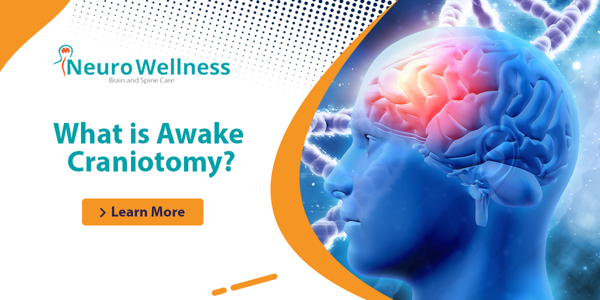
What is Awake Craniotomy?

What Is Awake Craniotomy?

Awake craniotomy, also known as awake brain surgery, is a specialized surgical procedure performed while the patient is awake and conscious. 

This advanced surgical technique allows neurosurgeons to monitor and protect critical brain functions during the operation. 

By engaging the patient during the procedure, surgeons can ensure the safe removal of brain tumors, epileptic foci, or other abnormalities without damaging essential brain areas responsible for speech, movement, and vision.

Conditions Treated with Awake Craniotomy

Some brain (neurological) diseases, such as some brain tumours or epileptic seizures uncontrolled by medications, DBS for Parkinson’s disease, surgery for spasticity, surgery for some psychiatric disorders not controlled by medication are treated via awake brain surgery.

Awake craniotomy is often used to treat complex neurological conditions, including:

  1. Brain Tumors: Particularly those located near eloquent brain areas, such as motor, language, or visual regions.
  2. Epilepsy: Cases where seizures are not controlled by medication and require precise localization of the epileptic focus.
  3. Deep Brain Stimulation (DBS): For treating conditions like Parkinson’s disease.
  4. Spasticity Surgery: In patients with severe muscle stiffness or spasms.
  5. Psychiatric Disorders: Cases resistant to conventional treatments.

Why Is Awake Craniotomy Advised?

The primary goal of an awake craniotomy is to preserve critical brain functions while treating the underlying neurological condition. Key areas targeted for protection include:

  • Motor Areas: Responsible for movement.
  • Language Areas: Critical for speech and communication.
  • Sensory Areas: Involved in processing sensory inputs like touch and vision.

This approach minimizes the risk of post-surgical complications, such as loss of speech, movement difficulties, or vision impairment.

The benefit of Awake Craniotomy.

Your comments will assist your surgeon in ensuring that the proper portion of your brain requires surgery. The surgery also reduces the chance of functional brain damage, which can impede your vision, mobility, and speech.

  • Enhanced Safety: Awake craniotomy ensures the surgeon avoids damaging critical brain regions.
  • Real-Time Feedback: The patient’s responses during surgery guide the surgeon in identifying functional areas of the brain.
  • Improved Outcomes: The risk of functional impairments is significantly reduced, enabling a faster recovery.
  • Precise Tumor Removal: Allows surgeons to maximize tumor resection while preserving neurological functions.
  • Reduction in Seizures: In cases of epilepsy, awake craniotomy often results in fewer or no seizures post-surgery.

Why Awake Craniotomy Is Performed

Doctors must ensure that they do not damage the eloquent part of the brain if a tumour or section of your brain that causes seizures needs to be surgically removed.

Prior to surgery, it’s tough to locate such locations precisely. Awake brain surgery helps the surgeon pinpoint which parts of your brain regulate particular functions, allowing them to avoid them.

How Is Awake Craniotomy Performed?

How Surgery is performed?

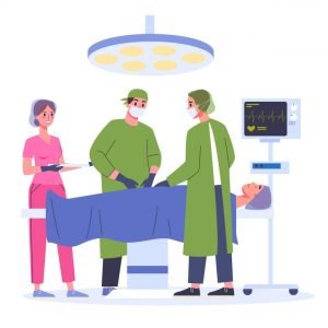
For sections of your awake brain surgery-Awake Craniotomy, the anesthesiologist will give you medicine to make you sleep. To guarantee your comfort, the neurosurgeon will administer numbing drugs to your scalp.

Doctors put your head in a set position during the surgery to keep it motionless and enhance surgical precision. Your hair will be trimmed in some places. After that, your surgeon will remove a portion of your skull to have access to your brain.

You’ll be given medication and asleep as a small portion of your skull is taken at the start of the procedure, as well as when physicians reattach it at the end of the procedure. Your anesthesiologist will cease giving you sedative drugs during surgery and enable you to wake up.

Your doctor will undertake brain mapping if your brain tumour or epileptic focal is close to parts of your brain that regulate vision, speech, or movement. This offers a map of the brain centre’s that govern each of these functions to your neurosurgeon. During surgery, your surgeon may be able to undertake brain mapping deeper in your brain.

This map is used by your neurosurgeon to avoid injuring certain locations and to keep their functions intact. Your surgeon can safely remove as much of your brain tumour or epileptic focus as feasible using brain mapping and 3-D computer pictures, reducing the risk of disrupting vital bodily processes.

Your medical team will also employ comprehensive 3-D computer scans of your brain, including intraoperative MRI and computer-assisted brain surgery, to guide the removal of as much of the brain tumour or epilepsy focal as feasible before and during surgery.

Anaesthesiologist and surgical team closely monitor and analyse your body and brain functioning, alerting your surgeon if the operation has a negative impact on your brain function.

Outcomes of Awake Craniotomy

  • For Epilepsy: Many patients experience a significant reduction in seizures post-surgery, with some achieving complete seizure freedom.
  •  
  • For Tumors: The procedure allows for maximum removal of the tumor while preserving vital brain functions. Additional treatments, like chemotherapy or radiation therapy, may be required for residual tumor cells.

Summary:

what is awake craniotomy

If you have awake brain surgery-Awake Craniotomy to treat epilepsy, you should notice a reduction in your seizures following the procedure. Some patients have no seizures at all, while others have fewer seizures than they did before the procedure.

Your neurosurgeon should have been able to remove the majority of the tumour if you had awake brain surgery to remove it. Other therapies, such as radiation therapy or chemotherapy, may be required to help eliminate any residual tumour cells.

FAQs About Awake Craniotomy

FAQs About Awake Craniotomy

1. What is awake craniotomy?
Awake craniotomy is a type of brain surgery performed while the patient is awake to preserve critical brain functions like speech and movement during the procedure.

2. Why is awake craniotomy necessary?
This surgery ensures that essential brain areas are not damaged during procedures like tumor removal or epilepsy treatment.

3. Is awake craniotomy painful?
No, numbing medications and careful monitoring by the anesthesiologist ensure the procedure is painless.

4. What conditions are treated with awake craniotomy?
Awake craniotomy is used to treat brain tumors, epilepsy, Parkinson’s disease, spasticity, and certain psychiatric disorders.

5. How long does recovery take after awake craniotomy?
Recovery varies by patient, but most individuals can return to normal activities within weeks, depending on the complexity of the surgery.

6. Are there risks associated with awake craniotomy?
As with any surgery, there are risks, including infection, bleeding, or neurological deficits. However, awake craniotomy minimizes these risks by preserving essential brain functions.

7. How do I prepare for awake craniotomy?
Your medical team will provide detailed instructions, including tests, fasting guidelines, and pre-surgical counseling to ensure you are mentally and physically prepared.

Minimally invasive pin hole surgery for narrowed brain vessel (MCA stenting) for brain stroke

Successful MCA Stenting for TIA in a 46-Year-Old Patient: A Case Study

A 46-year-old gentleman from North Karnataka presented with complaints of left-sided weakness and difficulty speaking for 10 days prior to his arrival at 1224, G-Floor, 26th Main, 9th block, Jayanagar, Opp to Ragiguddada Anjaneyaswamy Temple Arch, Bangalore, Karnataka 560069. On examination, he was conscious, alert, and oriented, with no noticeable motor or sensory deficits. Further investigation led to a diagnosis of a Transient Ischemic Attack (TIA), a condition often referred to as a mini-stroke. Here’s an in-depth look into his diagnosis, treatment, and recovery process.

MRI
Minimally invasive pin hole surgery f

Initial Diagnosis

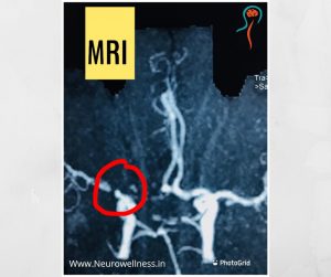
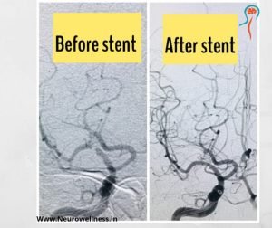
The patient underwent an MRI brain stroke protocol, which revealed watershed infarcts in the right middle cerebral artery (MCA) territory with high-grade stenosis of the MCA. To further evaluate the extent of the problem, a Digital Subtraction Angiogram (DSA) was performed, showing more than 95% focal stenosis, with significantly reduced blood flow into the distal branches of the M2 and M3 segments of the MCA.

This high-grade stenosis posed a significant risk of a major stroke, prompting the team to plan for MCA stenting after carefully explaining the benefits and risks to the patient.

What is MCA Stenting?

The Middle Cerebral Artery (MCA) is one of the major blood vessels supplying the brain. Stenting is a minimally invasive procedure that involves placing a small tube called a stent inside the narrowed artery to restore proper blood flow.

Unlike traditional brain surgery, MCA stenting doesn’t require opening the skull (craniotomy). Instead, thin wires and catheters, as small as a strand of hair, are inserted through a blood vessel in the thigh. These wires are guided all the way to the narrowed brain artery, where the stent is placed to open up the blocked area.


Procedure Overview

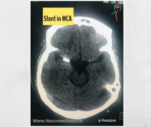
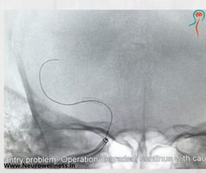
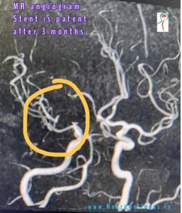
The patient was taken to the catheterization (Cath) lab for the procedure. A right femoral puncture was made to insert the guide wires and a micro-catheter. Using these tools, the team successfully reached the right MCA and placed a 2.25mm x 8mm stent in the brain vessel. The patient tolerated the procedure well, with no complications during or after the stenting.

Stenting
Brain Stroke

Post-Operative Results and Recovery

A post-operative CT scan showed no signs of bleeding or stroke. The patient experienced no complications and was able to walk the next day. He was discharged two days later, in good condition.

Stenting

Understanding Transient Ischemic Attacks (TIA)

A Transient Ischemic Attack (TIA), also called a mini-stroke, is a temporary blockage of blood flow to the brain. Unlike a major stroke, the symptoms of a TIA usually resolve within 24 hours. However, it is crucial to identify the underlying cause, as a TIA is often a warning sign of a more serious stroke.

Symptoms of TIA

  • Weakness in the face or limbs, typically on one side
  • Difficulty speaking or understanding speech
  • Dizziness or loss of balance
  • Temporary vision disturbances
  • Numbness or tingling sensations

CT scan

Diagnosing TIA

A variety of diagnostic tools are used to confirm a TIA and identify the cause:

  • Duplex scan
  • CT scan of the brain
  • MRI of the brain
  • MRA angiogram
  • Echocardiogram (ECHO)
  • Perfusion brain scan
  • DSA (Digital Subtraction Angiogram)

Importance of Early Detection and Treatment

TIAs are a critical warning sign, predicting a higher risk of a major stroke. Studies show that about 15% of people who experience a TIA will suffer a major stroke within three months. The risk increases to 30-40% within one year if left untreated. Therefore, prompt diagnosis and treatment are essential to prevent a future stroke.

CT scan

Preventive Treatments for Stroke

Several interventional procedures are available to prevent strokes in high-risk individuals, such as those who have experienced a TIA:

  • MCA Stenting: This is the procedure that was performed in this case to treat severe stenosis and restore blood flow to the brain.
  • Carotid Artery Stenting: This procedure is used to treat blockages in the carotid arteries in the neck, which supply blood to the brain.
  • Mechanical Thrombectomy: This is an emergency treatment used to remove large blood clots during an acute stroke.

These minimally invasive procedures can dramatically reduce the risk of a major stroke, especially in patients with a history of TIA.

Conclusion

In this case, the timely diagnosis of MCA stenosis and the successful MCA stenting procedure helped prevent a major stroke for the 46-year-old patient. With the growing availability of advanced interventional treatments, it is possible to reduce the risk of life-threatening strokes in patients with conditions like TIA.

For those at risk, early medical intervention can make all the difference in ensuring a full recovery. If you experience symptoms of a mini-stroke, it’s essential to seek medical attention promptly.


This surgery was performed, Consultant Neurosurgeon specializing in brain and spine surgery.

For more information or consultation, contact:
Email: ganeshneuros@gmail.com
Phone: 9845674662, 7349017701

FAQs

1.What is minimally invasive pinhole surgery for MCA stenting?

Minimally invasive pinhole surgery for MCA (Middle Cerebral Artery) stenting involves accessing the brain’s blood vessels through a small puncture, typically in the groin. Using specialized catheters and imaging techniques, a stent is placed in the MCA to restore proper blood flow.

2.What are the benefits of minimally invasive MCA stenting?

This approach offers several advantages, including reduced blood loss, shorter hospital stays, no need for large incisions, minimal scarring, and a quicker recovery compared to traditional open surgeries.

3.Who performs minimally invasive MCA stenting?

These procedures are conducted by specialists trained in interventional neurology, neurosurgery, or radiology, often referred to as endovascular surgeons. They undergo advanced training to master these delicate techniques.

4.What conditions can be treated with neuroendovascular surgery?

Neuroendovascular surgery can address various conditions, including brain aneurysms, acute ischemic strokes, arteriovenous malformations (AVMs), carotid artery stenosis, and carotid-cavernous fistulas.

5.What are the risks associated with MCA stenting?

While generally safe, potential risks include clot formation at the puncture site, allergic reactions to contrast dye, and the possibility of blood clots traveling to critical areas, which could lead to a stroke.

    DOWNLOAD EBOOK

      DOWNLOAD EBOOK

        DOWNLOAD EBOOK

          DOWNLOAD EBOOK

            DOWNLOAD EBOOK

              DOWNLOAD EBOOK