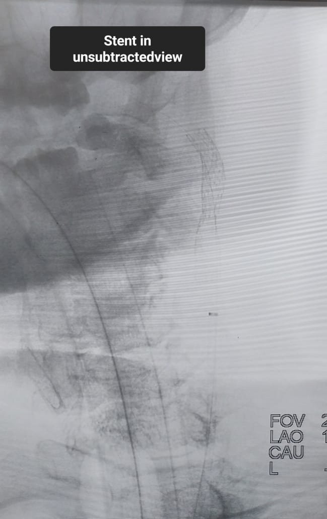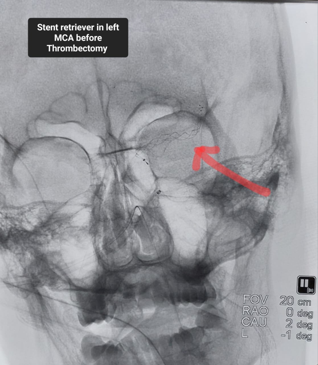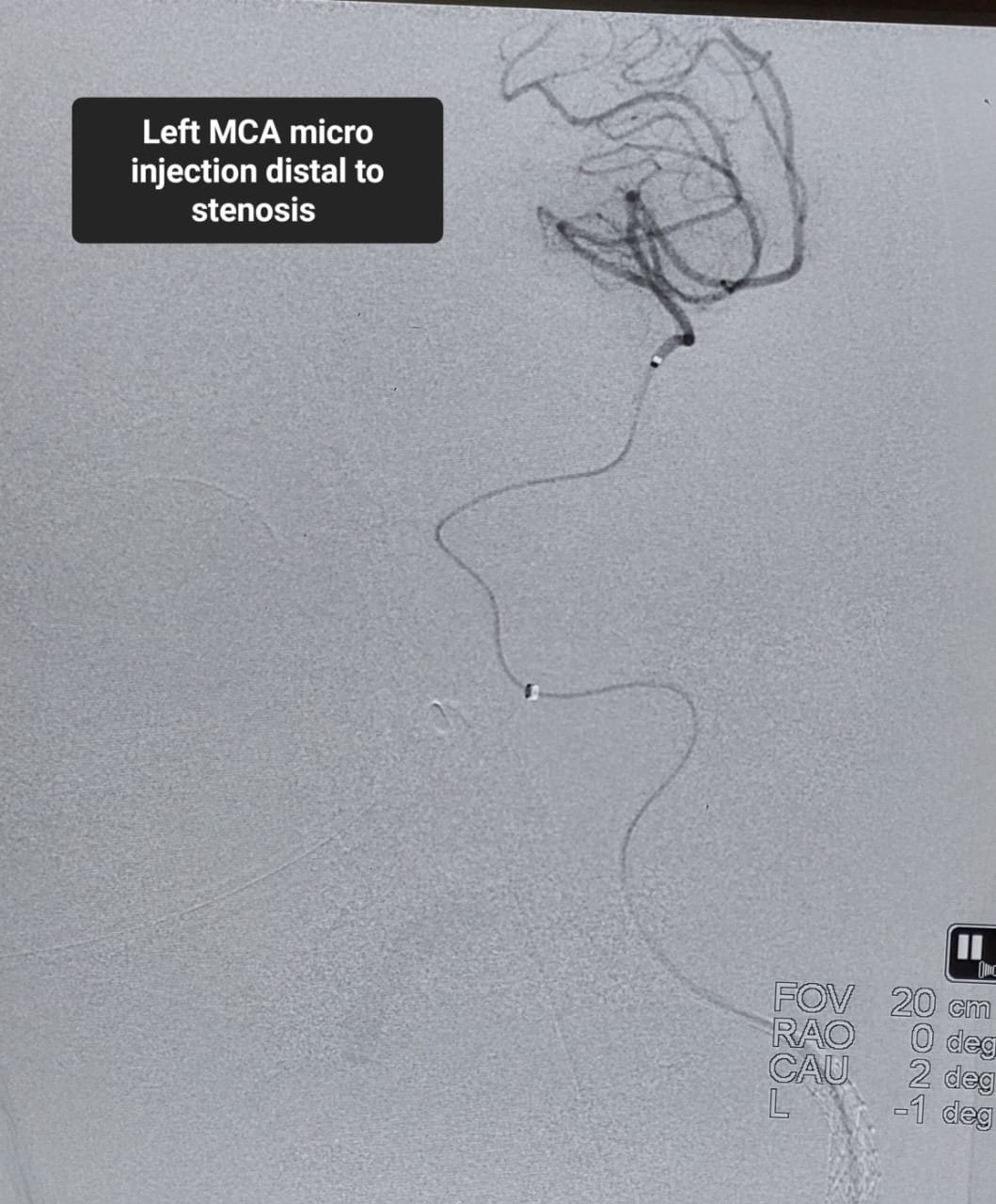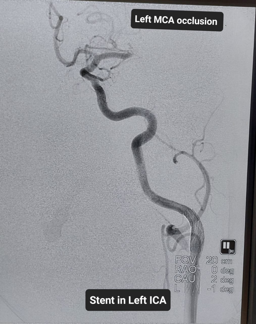
Part 1: A Stroke Emergency Caused by Two Major Arterial Blockages
Stroke can change a person’s life within minutes. Someone who was speaking and moving normally may suddenly develop facial weakness, paralysis of one side, speech difficulty, loss of vision, imbalance or reduced consciousness.
In an acute ischaemic stroke, an artery supplying the brain becomes blocked. Brain cells are deprived of oxygen and glucose, and potentially reversible brain dysfunction can progress to permanent injury unless blood flow is restored quickly.
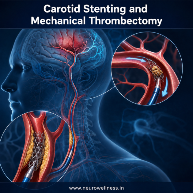
This NeuroWellness public-awareness case describes a 66-year-old man who presented approximately three hours after suddenly developing complete weakness of the right side. An urgent non-contrast CT scan did not show intracranial bleeding or a clearly established large infarction. Following rapid clinical and imaging assessment, intravenous thrombolysis was administered, and he was taken for cerebral angiography and possible mechanical thrombectomy.
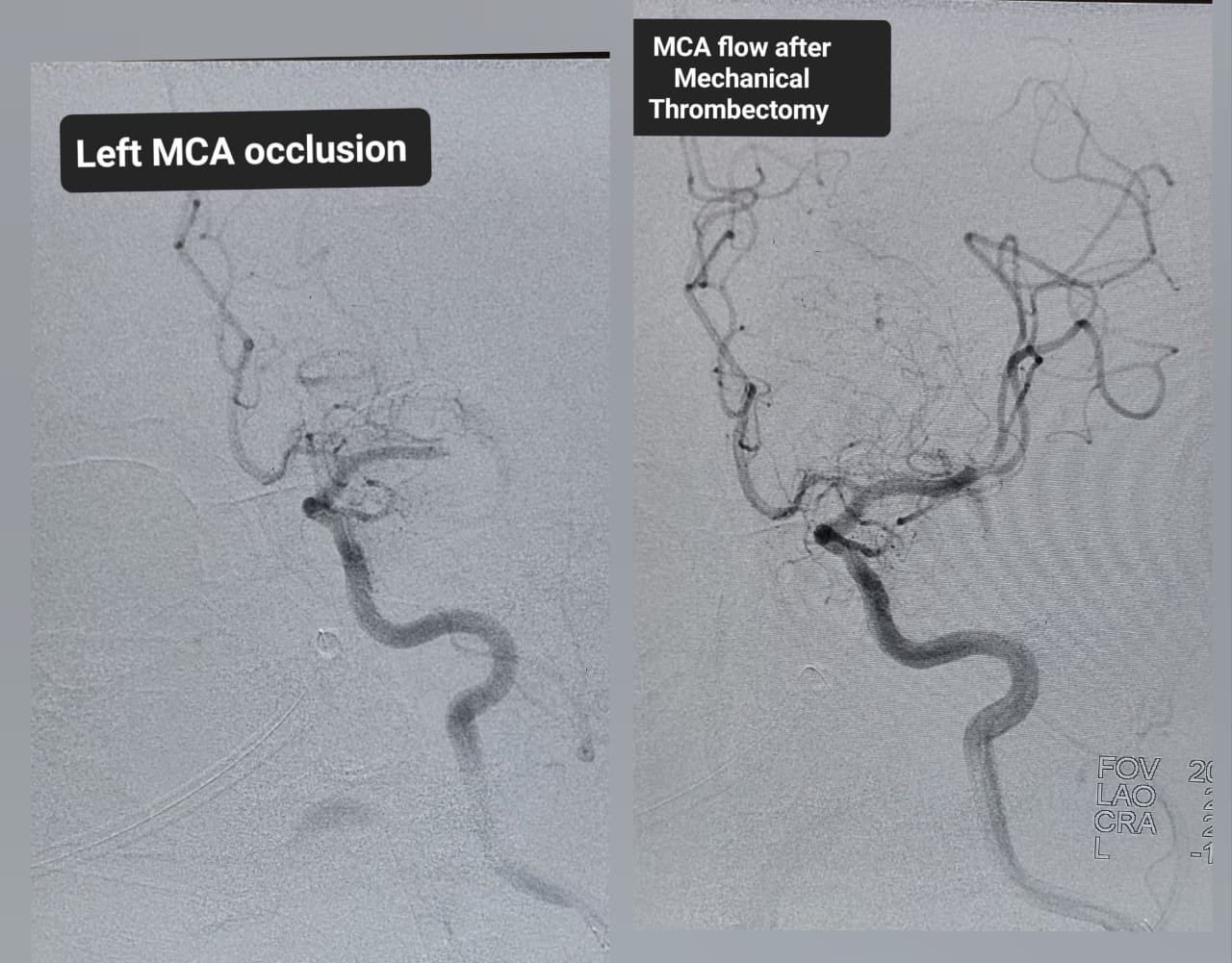
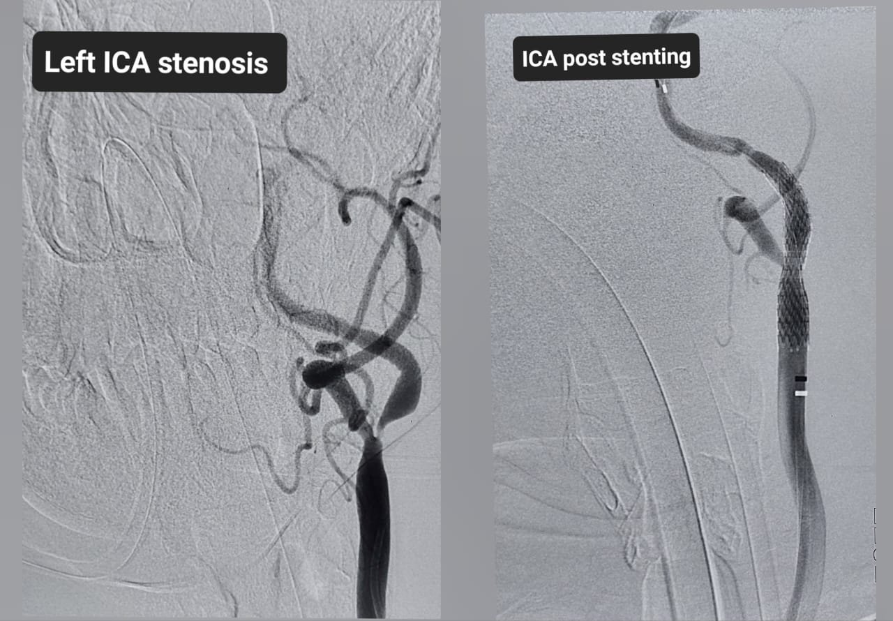
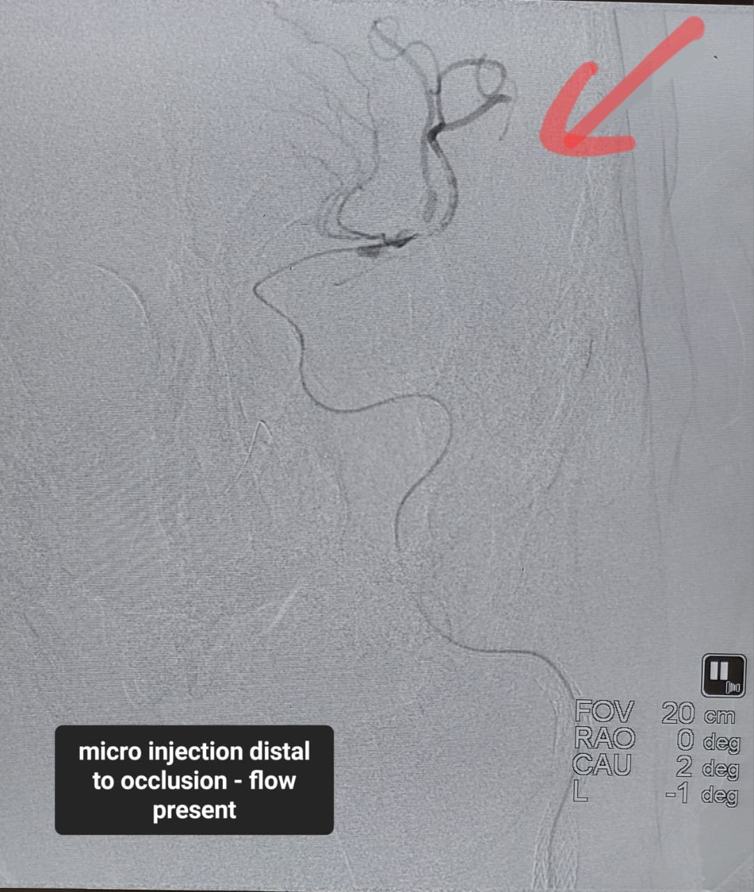
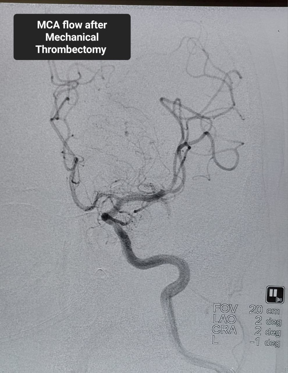
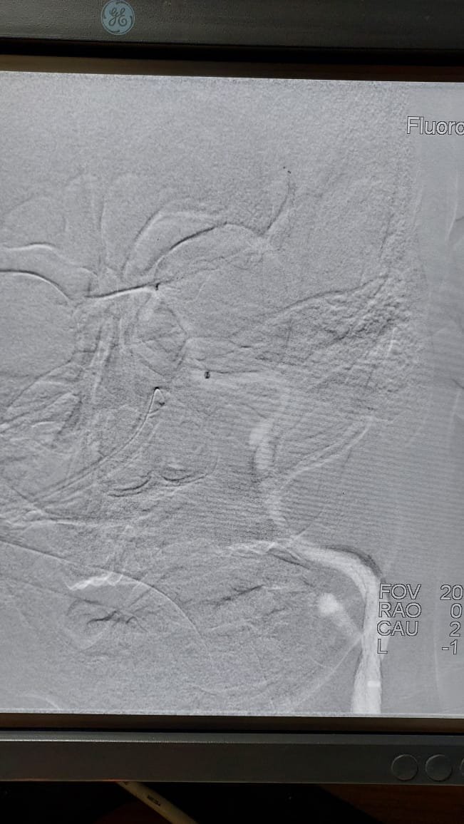
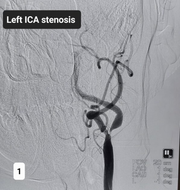
Angiography revealed approximately 95% stenosis at the origin of the left internal carotid artery together with absence of flow in the left middle cerebral artery. This meant that a critically narrowed neck artery and an intracranial large-vessel obstruction were threatening circulation to a major part of the left cerebral hemisphere.
The treating team performed carotid artery stenting to open the critically narrowed internal carotid artery and mechanical thrombectomy to remove the intracranial obstruction. By the following day, movement on the affected right side had improved to approximately Medical Research Council grade 2–3. This was encouraging, but long-term recovery would depend on the final extent of brain injury, durable reperfusion, complications, rehabilitation and the patient’s overall health.
The central lesson is not that every patient requires these procedures. It is that sudden stroke symptoms demand immediate transport to a stroke-ready hospital. Treatment is highly time-sensitive and must be individualised after expert assessment.
Recognising Stroke: Remember BE FAST
B — Balance: sudden dizziness, loss of balance, poor coordination or inability to walk normally.
E — Eyes: sudden loss of vision, blurred vision, double vision or a new visual-field defect.
F — Face: one side becomes weak, numb or droops while smiling.
A — Arm: sudden weakness or numbness of an arm, often accompanied by leg weakness on the same side.
S — Speech: slurred speech, inability to speak, wrong words or difficulty understanding language.
T — Time: note when the patient was last known to be normal and arrange emergency transport immediately.
Do not wait to see whether the symptoms settle. Do not give food, water, aspirin or home remedies unless directed by the treating team. Do not allow the patient to drive. In India, precious time may be lost by waiting at home or visiting multiple facilities without stroke imaging or intervention capability.
Why a Left-Brain Stroke Caused Right-Sided Paralysis
The left side of the brain controls movement on the right side of the body. A blockage in a major left-sided cerebral artery can therefore cause weakness or paralysis of the right face, arm and leg. A left-hemisphere stroke may also produce aphasia—the inability to speak, find words, understand language, read or write—even when consciousness is preserved.
Complete or near-complete loss of movement on one side is called hemiplegia. It may indicate a large vessel occlusion. Clinical examination establishes severity, while vascular imaging identifies the artery involved and helps determine whether endovascular treatment is appropriate.
Can the First CT Scan Be Normal During a Real Stroke?
Yes. A normal-looking early non-contrast CT does not exclude acute ischaemic stroke. During the first few hours, visible tissue changes may be subtle or absent. The immediate purpose of the scan is usually to exclude intracranial bleeding, detect a large established infarction and identify other structural abnormalities.
Treatment decisions are based on the complete picture: last-known-well time, neurological examination, blood glucose, blood pressure, medical history, medications, laboratory results and imaging. CT angiography or MR angiography can show an arterial blockage. Perfusion imaging may help selected patients by estimating irreversibly injured tissue and potentially salvageable brain.
Public takeaway: Sudden paralysis or speech difficulty remains a stroke emergency even when the initial plain CT does not show an obvious infarction.
Intravenous Thrombolysis
Intravenous thrombolysis uses a clot-dissolving medicine such as alteplase or tenecteplase in eligible patients with acute ischaemic stroke. It is commonly considered within 4.5 hours from last known well, although eligibility depends on clinical circumstances, imaging and current protocols.
The team must first exclude intracranial haemorrhage and assess contraindications such as active bleeding, particular recent operations or injuries, uncontrolled blood pressure, coagulation abnormalities and some medicines or illnesses. Thrombolysis can improve the possibility of recovery, but it can also cause serious bleeding.
For an eligible patient with a large vessel occlusion, thrombolysis may be administered even when thrombectomy is being planned. This is sometimes called bridging treatment. A large clot may not dissolve sufficiently with medication alone, making mechanical removal necessary.
Large Vessel Occlusion and Tandem Lesions
A large vessel occlusion blocks one of the major arteries supplying the brain, such as the internal carotid artery or proximal middle cerebral artery. It can cause profound weakness, gaze deviation, aphasia, neglect, visual loss or reduced consciousness.
The middle cerebral artery supplies extensive regions responsible for movement, sensation, language and attention. Absence of left middle cerebral artery flow therefore represents a dangerous interruption of circulation.
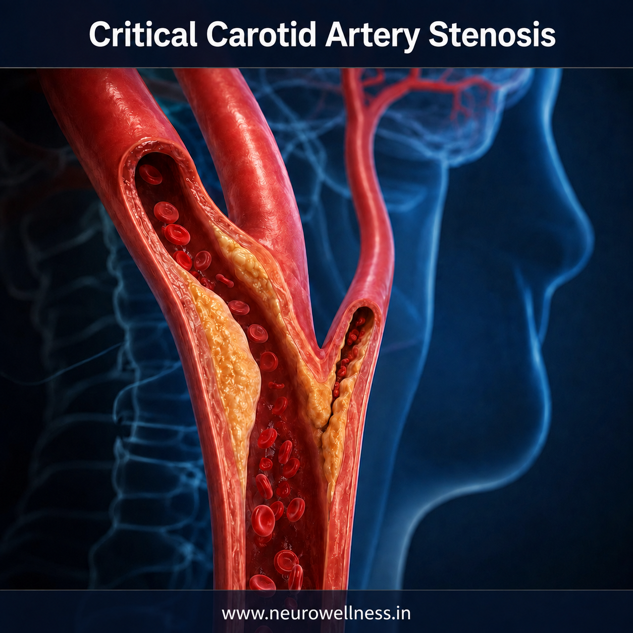
The combination of severe extracranial internal carotid disease and an intracranial obstruction is called a tandem lesion. Atherosclerotic plaque may critically narrow the carotid origin and release clot or plaque material that travels upwards. A 95% stenosis leaves a very narrow channel and may also make access to the intracranial clot technically difficult.
What Is Carotid Artery Stenting?
Carotid stenting is an endovascular procedure used in selected patients to open a narrowed carotid artery. A catheter is advanced under X-ray guidance, angioplasty may widen the channel, and a self-expanding stent is positioned across the stenosis to support the artery and improve flow.
During an acute tandem-lesion stroke, stenting may also create a pathway for devices to reach the intracranial clot. It is not automatically required for every patient. Risks include embolisation, vessel injury, bleeding, stent thrombosis, reperfusion injury, blood-pressure or heart-rate changes, contrast-related kidney injury and later restenosis.
Learn more : carotid artery stenosis / carotid stenting
What Is Mechanical Thrombectomy?
Mechanical thrombectomy is a minimally invasive catheter procedure that physically removes a clot from a major brain artery. A stent retriever may engage the clot, aspiration may suction it out, or both methods may be combined. The objective is reperfusion—restoration of blood flow to threatened brain tissue.
Thrombectomy is strongly time-dependent. It is usually performed as soon as possible in eligible large vessel occlusion strokes. Carefully selected patients may benefit up to 24 hours after last known well when clinical and advanced imaging criteria show salvageable tissue. The longer window must never be interpreted as permission to wait.
Eligibility depends on the blocked artery, severity, established infarct volume, remaining viable tissue, pre-stroke function, bleeding risk, comorbidities and local protocols.
Why Both Procedures Were Needed
This emergency involved two connected problems: a critically narrowed gateway artery in the neck and an occluded major artery inside the brain. Treating only the intracranial clot might leave access or durable flow compromised in some anatomies. Treating only the carotid stenosis would not necessarily remove the clot already obstructing the middle cerebral artery.
A combined strategy was therefore used—carotid stenting to open the severely narrowed left internal carotid artery and mechanical thrombectomy to restore left middle cerebral artery flow. The order and exact technique for tandem lesions vary; this case should not be treated as a universal procedural formula.
Part 2: Recovery, Rehabilitation and Stroke Prevention
What Does Grade 2–3 Power Mean?
Muscle strength is commonly recorded on the Medical Research Council scale. Grade 0 means no contraction; grade 1 is a flicker; grade 2 is movement when gravity is eliminated; grade 3 is movement against gravity but not resistance; grade 4 is movement against resistance; and grade 5 is normal strength.
Improvement from hemiplegia to grade 2–3 movement by the next day can be a positive early sign. It suggests that some motor pathways are functioning after reperfusion. However, one examination cannot predict final outcome. Strength may improve further, remain limited or worsen because of infarct evolution, swelling, bleeding, re-occlusion, infection, seizures or other complications.
The First 24–72 Hours
After thrombolysis, thrombectomy and carotid treatment, patients are usually observed in a dedicated stroke unit or intensive-care environment according to severity.
Repeated neurological examinations to detect deterioration.
Careful blood-pressure, oxygen, glucose, temperature and heart-rhythm monitoring.
Follow-up imaging to assess infarction and exclude haemorrhage.
Access-site and peripheral-circulation assessment.
Swallow screening before oral food, water or medicines.
Prevention of aspiration, pressure injury, deep-vein thrombosis and infection.
Individualised antiplatelet planning when a carotid stent has been placed.
Investigation of stroke mechanism and early rehabilitation assessment.
Antiplatelet treatment helps prevent clot formation inside a carotid stent, but recent infarction and thrombolysis may increase bleeding risk. Patients must never add, stop or change aspirin, clopidogrel, ticagrelor, anticoagulants or other blood-thinning medicines without instructions from the stroke team.
Reperfusion Is the Beginning, Not the End
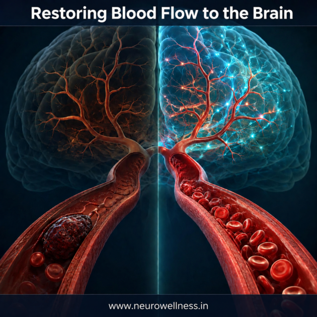
Opening an artery can save threatened tissue, but it does not instantly reverse every effect of stroke. Some brain tissue may already be injured. Recovery depends on infarct size and location, time to reperfusion, collateral circulation, complications, age, overall health, cognition, mood and rehabilitation.
Rehabilitation should begin when the patient is medically stable. Physiotherapy addresses positioning, balance, standing, walking and strength. Occupational therapy helps relearn dressing, bathing, eating and use of the affected hand. Speech and language therapy addresses aphasia, slurred speech and swallowing problems. Cognitive and psychological support may be needed for memory, attention, anxiety, depression or emotional change.
Families should not force an unstable patient to walk or pull a weak arm, which can injure the shoulder. Food or water before swallow clearance can enter the lungs. Rehabilitation must be progressive, supervised and goal-based.
Preventing Another Stroke
An acute stroke is also a warning that underlying vascular disease requires long-term treatment. Secondary prevention may include prescribed antiplatelet therapy, intensive lipid lowering, blood-pressure control, diabetes management, tobacco cessation, healthy weight, appropriate physical activity and follow-up vascular imaging.
Doctors may evaluate heart rhythm for atrial fibrillation, perform echocardiography when appropriate and assess other brain-supplying arteries. Even when carotid atherosclerosis appears to explain the event, a complete work-up can reveal additional treatable causes.
Control high blood pressure, diabetes and LDL cholesterol.
Avoid cigarettes, beedis and smokeless tobacco completely.
Treat atrial fibrillation and other heart disease as advised.
Maintain appropriate activity, nutrition, weight and sleep.
Attend neurological and carotid-stent follow-up.
Continue prescribed medicines even when strength and speech improve.
Stroke Awareness Challenges Across India
India has metropolitan, semi-urban and rural communities with major differences in access to CT imaging, stroke units and neurointervention. Delays may arise from failure to recognise symptoms, waiting for relatives, attempting home remedies, visiting multiple facilities, arranging transport or not communicating with the receiving hospital.
Families can prepare by learning BE FAST, identifying a nearby hospital with 24-hour CT and stroke evaluation, knowing a regional thrombectomy centre, keeping an updated medication list and recording the last-known-well time. Calling ahead during transport can help activate the hospital stroke pathway.
A patient who wakes with weakness may have been last known well when going to sleep. Advanced imaging can still identify some candidates for reperfusion treatment. Therefore, no family should assume that a patient is automatically outside the treatment window.
Common Myths That Cause Delay
Myth — “The patient is conscious, so it cannot be a major stroke.”
Fact — A disabling large vessel occlusion can occur while consciousness is preserved.
Myth — “The first CT is normal, so there is no stroke.”
Fact — Early ischaemic stroke may not be visible on plain CT.
Myth — “We should wait for our regular doctor.”
Fact — The patient should proceed immediately to emergency stroke assessment.
Myth — “Thrombolysis and thrombectomy are the same.”
Fact — Thrombolysis is clot-dissolving medication; thrombectomy physically retrieves or aspirates a large clot.
Myth — “Thrombectomy can be done for every stroke.”
Fact — It is used for selected arterial occlusions after clinical and imaging assessment.
Myth — “If weakness improves, rehabilitation is unnecessary.”
Fact — Fine hand function, balance, language, swallowing, cognition, mood and endurance may still require therapy.
Questions Families Should Ask
Was the stroke ischaemic or haemorrhagic, and which artery was blocked?
Was blood flow restored, and what does follow-up imaging show?
Was a carotid stent inserted, and which medicines are essential for it?
Is swallowing safe, and when should rehabilitation begin?
What caused the stroke and which investigations remain pending?
What are the blood-pressure, glucose and cholesterol targets?
When is the next clinical and vascular-imaging follow-up?
Which warning signs require immediate reassessment?
When to Seek Emergency Help After Discharge
Urgent reassessment is required for new or worsening facial droop, arm or leg weakness, speech difficulty, loss of vision, severe unusual headache, repeated vomiting, seizure, reduced consciousness, chest pain or breathing difficulty. Bleeding, black stools, vomiting blood or rapidly increasing swelling at the catheter access site also require prompt attention.
Recurrent symptoms may be brief. Even if weakness or speech difficulty resolves within minutes, it may represent a transient ischaemic attack or another stroke and must not be ignored.
Key Lessons From This Case
This anonymised case illustrates that sudden right hemiplegia is a medical emergency; an early CT may exclude haemorrhage without yet showing a large infarction; and rapid vascular assessment can reveal a complex tandem lesion. Here, critical left internal carotid stenosis and absent left middle cerebral artery flow were addressed using carotid stenting and mechanical thrombectomy after thrombolysis.
Early improvement to grade 2–3 right-sided movement was encouraging, but it remains an individual observation rather than a guarantee. Stroke treatment requires rapid, individualised decisions by an experienced multidisciplinary team.
NeuroWellness public-awareness message: Stroke is a brain emergency. At the first sign of sudden weakness, facial drooping, speech difficulty, visual change or loss of balance, seek emergency medical care immediately. Record the last-known-well time. Do not wait for symptoms to improve.
Frequently Asked Questions
1. Can stroke occur despite a normal CT scan?
Yes. Early ischaemic changes may not be visible on non-contrast CT. Doctors combine the scan with examination and vascular imaging.
2. What is the usual thrombolysis window?
It is commonly considered within 4.5 hours of last known well in eligible patients and should be delivered as early as safely possible.
3. What is the thrombectomy window?
It should be performed as soon as possible. Selected patients may benefit up to 24 hours based on clinical and advanced imaging criteria.
4. Why might a carotid stent be needed?
Severe carotid stenosis may impair brain blood flow or obstruct access to the intracranial clot. Stenting is an individual specialist decision.
5. Does early return of movement guarantee complete recovery?
No. Early improvement is encouraging, but monitoring, rehabilitation and secondary prevention remain essential.
6. Can stroke be prevented?
Not every stroke is preventable, but controlling vascular risk factors, avoiding tobacco and taking prescribed medicines can substantially reduce risk.
References
- American Stroke Association. Ischemic Stroke (Clots): medical therapies and mechanical clot removal.
- American Stroke Association. Acute Ischaemic Stroke Infographic: BE FAST and time-sensitive reperfusion treatment.
- American Heart Association/American Stroke Association. Guidelines and resources for early management of acute ischaemic stroke.
Medical disclaimer: This article is intended for public education. It does not replace examination, diagnosis or treatment by a qualified medical professional. Treatment eligibility, procedural strategy, medication and recovery differ between patients.
Author and procedure performed by Dr. Ganesh Veerabhadraiah
Dr. Ganesh Veerabhadraiah
Consultant – Neurosurgeon, Neurointerventional Surgery, Spine Surgeon (Neuro)
23+ Years Experience Overall (17+ years as Neuro Specialist)
Available for Consultation: Jayanagar 9th Block & Kauvery Hospital, Electronic City